What is a Visual Field Test and why it's important

      Perimetry goes by many names including: visual field (VF) testing, para-central  and/or peripheral vision testing, field of vision testing, amongst others. Similar to central visual acuity (commonly referred to as 20/20 or 6/6), perimetry examines how wide the eye can see, known as peripheral vision. When retinal cells, optic nerve fibers, or visual pathways in the brain are damaged, patients experience missing areas in their field of vision known as a visual field defect, scotoma, or blind zone.
      Was erkennt der Gesichtsfeldtest?

      What can a visual field test detect?

      Perimetry results are used to diagnose and monitor central, paracentral and peripheral vision loss. Testing is generally performed monocularly, but situations do arise where binocular testing is warranted, such as driving requirements. Analysis of perimetry data (VF charts) is useful in a myriad of ocular, systemic, and neurological conditions including – but not limited to – glaucoma, cone-rod dystrophy, retinitis pigmentosa, macular degeneration, optic neuritis, congenital optic neuropathies, brain tumors, lesions in the brain due to strokes, pituitary disorders, multiple sclerosis, brain trauma, and many more.

      When VF testing is required:

      • You are diagnosed with a progressive eye condition which requires regular monitoring (such as glaucoma or retinitis pigmentosa)
      • You are experiencing any concerning changes in your vision
      • You feel that your peripheral vision may be worsening (perhaps from blind spots or missing spots in your vision)
      • You find that you are constantly bumping into walls/objects or getting lost in crowded places (possibly because your side vision is restricted or limited)
      • You notice while reading that you are missing words or losing your place frequently
      • You know your vision is off, but you can’t describe why exactly 
      • You must renew your driver’s license and need the mandatory visual field testing

      When VF testing is recommended: 

      • To assess the severity, type, and progression of vision loss for patients who have suffered brain damage due to stroke, trauma, or tumors
      • To monitor vision loss for (likely) non-progressive eye conditions such as past ischemia or traumatic optic nerve damage
      • To evaluate if your multiple sclerosis (with relapses) is  affecting your vision and treat if necessary

      When you can spare on VF tests

      • If you vision problems are due to cataract or refractive problems (according to your eye care provider) 
      • If your visual complaints do not relate to optic nerve or retinal damage (must be confirmed by your eye care provider)
      • If your visual field limitations result from a droopy eyelid (and your eye care provider states no other problems exist)

      Find out if you are a good candidate

      Instructions for testing the visual field. Fedorov Restore Vision Clinic Berlin

      VF Testing Procedure

      Perimetry Testing Instructions . When performing perimetry, careful instructions must be given to the patient to establish accurate and reliable results. Patient instructions significantly affect perimetry results. Usually, one eye will be covered while the other eye is focused on a central target using a pre-calculated lens power. The patient’s chin should be on the chinrest and forehead against the forehead bar. The technician should inform the patient to only look at the central target during the entirety of the test and to only press the button on the handheld controller when the patient is sure he/she saw a flashing light.


      Gesichtsfeldtestverfahren. Eine Untersuchung der blinde Zonen

      The technician should properly align the eye along the center of the pupil and monitor central fixation of the eye during the entirety of the testing. The technician can also remind patients to continue only looking straight ahead if needed and inform patients of the approximate remaining test time. If the patient becomes fatigued during the testing, the assessment can be temporarily paused until the patient is ready to continue. Proper instructions and technician training can improve reliability of perimetry results thus enhancing patient care and clinic efficiency.

      Six major challenges while doing VF test

      As with all diagnostic testing, high quality and reliable results are critical. Unlike many diagnostic machines perimetry relies on subjective responses.

      1

      Being confused by instructions

      2

      Unfamiliar with test procedure

      3

      Struggling to keep fixation

      4

      Fatigue & daydreaming

      5

      Dread of visual field testing

      6

      Severe glare or light sensitivity

      Unlike many ocular diagnostic apparatuses that mostly only need proper scan alignment and high signal quality, perimetry relies on subjective responses. Patient responses are often influenced by biases due to the patient wishing to score well, being confused by the instructions, or being unfamiliar with how the test functions. Such variables may lead to false positive and false negative responses. Additionally, subjects oftentimes struggle to stay fixated on the central point, and instead, reposition their gaze toward the light stimuli – this is known as a fixation loss. Furthermore, patients often dread visual field testing due to the boring and mentally taxing nature of the test itself. Patient fatigue, distractions, daydreaming, and even falling asleep are frequently challenges technicians face when performing perimetry with patients. Such occurrences yield poor subjective responses making the perimetry test results unreliable. False positives, false negatives, fixation losses can also invalidate perimetry results. Before data interpretation, ensuring the results are reliable is paramount. If the results are deemed unreliable, perimetry testing should be repeated. Additionally, the standard of care is to validate perimetry results with several tests performed on different days. Oftentimes glaucoma treatment is not even initiated until visual field defects are confirmed after several consistent and reproducible visual field defects. All of this highlights the challenges clinicians face when conducting perimetry in clinics.

      Is a visual field test necessary?

      Although this is not a test that patients typically enjoy, visual field testing is the worldwide standard of care for assessing known or suspected vision loss. It is also used in diagnosing and monitoring the severity and progression of conditions like glaucoma.

      How can I tell if I have visual field loss?

      People may notice visual field loss by missing vision in one or both eyes, not seeing off to the side very well, bumping or tripping on objects, not noticing someone until they are right in front of you, or even trouble with reading or writing. Visual field loss is sometimes difficult to self-diagnose due to the brain “filling in” missing spots of vision. If you ever suspect visual field loss, the safest thing to do is get a visual field test.

      What does Visual Field Testing detect?

      Visual field testing looks for areas of your vision that aren’t seeing as well as they should. This could be your side vision, the top of your vision, the bottom of your vision, the vision around your blind spot, or even the center of your vision. Areas of vision correspond with areas of the retina and optic nerve. So, by knowing where vision loss is, your eye care provider can know which part of the retina or optic nerve may be unhealthy.

      What is required of you during this test?

      You need to keep looking straight at the center target directly in front of you. The machine will give off small flashes of light. When the flashes happen, press the button on your handheld device. Do not search for the flashes – let them come to you. The lights will be in different locations and may have different levels of brightness. Sometimes the light will be too dim for you to see – that is okay. Blinking is fine, but if your eyes start to feel dry, the test can be paused until you are ready to start again. Just relax and don’t try too hard.

      How reliable is a visual field test? Is it accurate?

      When performed correctly, visual field tests are very accurate and sensitive at detecting visual field loss. Overall, visual field tests are reliable 52% of the time. The professional interpreting the test can easily tell if the test is reliable or if the test must be repeated. The most common reason a test is unreliable is if the test-taker stops looking at the center target. When thorough and proper instructions are given to the test-taker reliability and accuracy greatly improves.

      What does a normal visual field test look like?

      For each eye, a normal visual field test extends about 100° toward your ears, 60° toward your nose, 60° up, and 70° down. Your “blind spot” is located about 15° toward your ears in each eye.

      Reports say that results are outside normal limits. What does it mean?

      First, results of several visual field tests would need to say “outside normal limits” for this to be verified. If verified, this means that you are seeing worse than normal people of your age. This could happen for many reasons. Ask us or your eye care provider for further clarification if you have questions.

      Can a visual field test be wrong?

      Because visual field testing requires careful attention and quick responses from the test-taker, good results can sometimes be difficult to obtain. False positives and false negatives as well as fixation loss may invalidate results. Sometimes the test-taker may get distracted or tired during the test also. For these reasons, multiple tests from different days are oftentimes needed to accurately determine the presence and extent of vision loss.

      How does Visual Field Testing impact driver’s license requirements?

      Both peripheral and central vision are used when driving, with peripheral vision being arguably more important. Countries and states differ on the minimum acceptable vision needed to be generally regarded as safe for driving. Eye care practitioners can fill out necessary forms and provide expert guidance on driving eligibility and safety regarding vision.

      Read more how Fedorov Restoration Therapy can help regain your drive licence back

      Are Visual Field Testing covered by medicare insurance?

      Visual field testing is generally fully – or at least partially – covered by insurance as long as you are suspected of having vision loss, in which case, visual field testing is medically necessary.

      Alle Fotos - 1 von 1 (5)

      Not sure if you need
      Visual Field Testing?


      Book An Appoitment For Your Evaluation

      Importe - 1 von 1 (6)

      Glaucoma and VF testing

      For decades, the most widely used diagnostic instrument for glaucoma management has been the visual field test. While other diagnostic assessments may demonstrate and quantify tissue loss or other structural changes, visual field testing is perhaps more clinically relevant as this evaluates the actual vision loss that the patient is experiencing. Due to its progressive nature, glaucoma requires constant monitoring to ensure vision loss is not continuing. VF testing is generally considered to be the most important assessment when managing glaucoma.

      For patients newly diagnosed, several visual fields are conducted in the first year to establish baseline measurements. Frequency of visual fields after that depend on the rate of progression for each patient, but generally are performed 1-3 times per year. If glaucomatous changes are observed, the frequency of visual field testing may increase. If the glaucoma is stable, the frequency of visual field testing may decrease. Visual field testing is critical for all patients with glaucoma as this data informs and guides clinicians on the best course of treatment to preserve vision.

      How many fields does it take to detect a deteriorating with certainty?

      Wenn Gesichtsfeldtests erforderlich sind?

      For the vast majority of glaucoma patients, vision loss occurs slowly, although some forms of glaucoma have a more aggressive nature. Generally 3-5 visual fields are needed to establish a baseline understanding of the field of vision. Once these initial tests are complete, your eye care provider will know the exact extent of your glaucoma or vision loss and be able to decide the best treatment protocol for you. Visual field testing is then generally performed 1-3 times per year to monitor for changes. The more severe the glaucoma, the more often visual field testing is needed.

      Also, if changes are observed with visual field testing, then testing may need to occur more often. Visual fields provide useful data which can demonstrate lack of reliability (the visual field test must be repeated), stable vision, or progressive vision loss. The more visual fields performed the more information your eye care provider will have to make the best decisions regarding your glaucoma treatment.

      How to read Visual Field Testing?

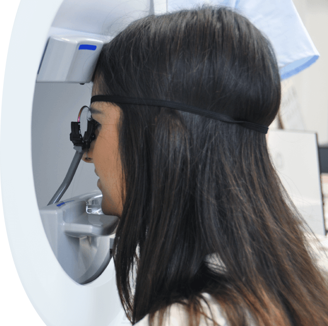
      Perimetry Test Results. Unlike central vision acuity which is measured by what letter size can be seen from a given distance, visual field testing measures the threshold of vision in decibels (dB). A smaller distinguishable stimulus threshold equates to more sensitive vision detection. These results are depicted using dB numbers for the area of peripheral vision being measured. The dimmer the light stimulus visible to the patient, the more sensitive the vision is for that particular point in the field of vision. Perimetry usually tests dozens of different visual points. Here are common ways the data is analyzed and depicted.

      Numerical plot: Displays the threshold for all visual points tested.

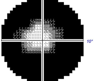
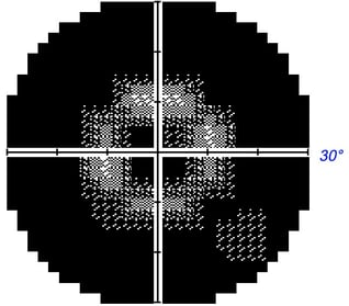
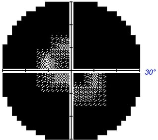
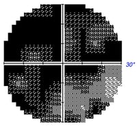
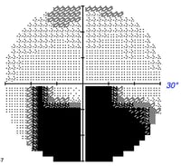
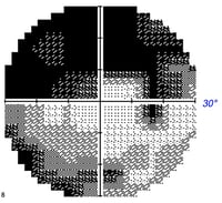
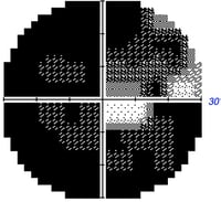
      Grayscale plot: Graphically depicts visual field loss. Normal areas are white and affected areas are grey/black. The level of darkness denotes the magnitude of vision loss for that area with darker being more severe. 

      Total deviation plot: Compares the patient’s results with normal age-matched controls and graphs areas of potential vision loss. This data can be influenced by extraneous factors though (ex. cataracts). 

      Pattern deviation plot: Similar to the total deviation plot except this is adjusted for any generalized depression seen on the total deviation plot. This is helpful with conditions affecting all visual points (ex. cataracts). 

      Mean deviation: An average of the results from all tested locations and compared with normal values.  

      Pattern standard deviation: Provides data about localized rather than generalized visual field loss.  

      Hemifield test: Compares visual field loss of the upper hemifield to the lower hemifield. This is especially useful with glaucoma suspects. 

      Many forms of visual field defects exist corresponding with different ocular and neurological conditions. For instance, a bitemporal hemianopia is associated with a pituitary gland tumor of the optic chiasm while an enlarged blind spot is an indicator of optic neuritis.

      Perimetry Interpretation. As with all medical diagnostic assessments, the data interpretation and disease management need to be determined by a trained professional. Having said that, there are some basic fundamentals non-professional may use to roughly interpret perimetry results. Before testing should be interpreted, one must ensure the test results are reliable (discussed in greater detail in the next section). 

      Firstly, each eye should have a blind spot that corresponds with the optic nerve head as this area of the eye does not detect light, but carries information to the brain from other areas of the retina. If the blindspot is missing, the contralateral eye may not have been appropriately covered. 

      The gray scale plot (usually at the top) is a way to quickly visualize and generally assess peripheral vision loss. Though the gray scale is a great starting point, it is not always accurate. The gray scale also reveals which eye has been evaluated – if the blind spot is on the left then the left eye was evaluated; if the blind spot is on the right the right eye was evaluated. 

      The mean deviation analyzes and quantifies if the eye has above average sensitivity (positive number), average sensitivity (zero), or reduced sensitivity (negative number). The more negative the number, the greater the overall vision loss for that eye. A high pattern standard deviation number reveals if there is a significant difference between the upper and lower hemifield, which is especially useful in glaucoma evaluation. This can easily be interpreted by looking at the glaucoma hemifield test (GHT) that simply states if this is within normal limits, borderline, or outside normal limits.  

      The total deviation plot displays the total magnitude and location of vision loss; remember this can be affected by media opacities (cataracts, corneal scars, vitreous hemorrhages, etc). Usually considered more useful is the pattern standard deviation plot, which displays the vision loss after taking into account the total deviation.

      Examples of Visual Field Testing 

      I. Optic Nerve Damage / Atrophy

      Normal Field of Vision
      Normal Field of Vision Restore Vision Clinic Berlin
       
      Dominant Optic Atrophy
      Importe - 1 von 1 (12)
      Ischemic Optic Neuropathy
      Importe - 1 von 1 (13)
      Severe Optic Neuritis
      Importe - 4 von 4
      Mild 
      Glaucoma 
      Importe - 3 von 4
      Advanced
      Glaucoma
      Importe - 2 von 4
      Very advanced Glaucoma
      Importe - 1 von 4
      Leber Optic Atrophy
      Importe - 1 von 1 (15)
       

      II. Retinal Dystrophies

      Mild Retinitis Pigmentosa
      Importe - 1 von 1 (16)


      Late Retinitis Pigmentosa
      Importe - 1 von 2


      Cone-Rod Dystrophy
      Importe - 5 von 6


      End Stage
      of RP
      Importe - 6 von 6

      Get in touch today