.jpeg)

Vision necessary for orientation and mobility is particularly dependent on visualizing low-contrast objects such as curbs, steps, and shadows. Driving presents additional low contrast challenges during rain, dusk, fog, snow, and at night. Even typically simple tasks, like reading a paper book, can quickly become visually difficult if contrast is reduced (such as with poor lighting or if the text is faint). Contrast sensitivity significantly contributes to overall visual quality and partially explains why a person seeing “20/20” may still be afflicted by poor vision.
From a patient's point of view, poor contrast sensitivity means constantly having foggy, hazy, or cloudy vision. When severe, it greatly affects night driving, visualizing road signs, seeing pedestrians in the dark, and even increases the risk of falling (especially if the person fails to see the step below).

Interestingly, human vision operates better under a wide range of luminances than contrasts, which is why humans are able to see both during the day and night (although this vision may be limited by contrast sensitivity). Human eyes can both light-adapt and dark-adapt depending on the overall luminance. However, in bright and dim lighting situations humans sometimes struggle with contrast or the ability to perceive an object from a similar appearing background. Another way to think about contrast and luminance is when editing photos. If you have ever edited photos or altered computer display settings, you were likely altering the brightness (similar to luminance) and contrast.

The point at which an object becomes indistinguishable from its background is the contrast threshold. Humans can visualize the contrast threshold during sunsets or during a foggy or rainy day. Contrast sensitivity varies by person depending on age, cataracts, ocular health (ex. glaucoma, diabetes, retinal dystrophies, retinal degenerations), and visual cortex processing. Loss of contrast sensitivity oftentimes precedes visual field defects and visual acuity deterioration in cases of disease. This is especially seen with glaucoma, diabetic retinopathy, and cataracts. Inherited retinal diseases are also known to significantly affect contrast sensitivity. The most common cause of diminished contrast sensitivity is normal aging of the retina.
Clinicians measure contrast sensitivity using a series of sine-wave gratings of different sizes, luminances, and intensities. Thick bars represent low spatial frequencies, while thin bars represent high spatial frequencies – similar to high and low pitches in a hearing examination. Low Contrast Visual Acuity (LCVA) combines contrast sensitivity testing with visual acuity by actually measuring visual acuity in a low-contrast setting. This method of visual assessment is a far more accurate assessment of visual function than traditional visual acuity. This form of visual assessment is beneficial in determining the true visual function in patients and is likely a better predictor of visual quality than visual acuity alone. In fact, contrast sensitivity testing is arguably the most relevant clinical vision test when evaluating “real world vision” as daily life presents a wide range of high and low contrast situations. Previously, contrast sensitivity testing and LCVA were rarely performed due to clinicians believing that contrast sensitivity loss was untreatable. However, recent evidence suggests improvement in contrast sensitivity is attainable.
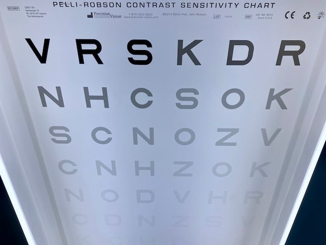
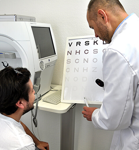
The most commonly used tests to assess contrast sensitivity are Test Bailey-Lovie Chart or Regan (Australia) and Pelli-Robson Contrast Test (USA). The Pelli-Robson Contrast Sensitivity Chart (PR) displays six 20/60 size letters on each row. These letters gradually become less contrasted against the white background as you move down the chart; high contrast letters are at the top and low contrast letters are at the bottom. There are three letters per group. Each time contrast is reduced, the next group of three letters is harder to distinguish. Patients begin from the top and proceed downward until they can no longer discern the letters. A score is determined by the last group in which a person could correctly identify the majority of the letters. Scores, a number and decimal, are measured by log contrast sensitivity. PR scores are arranged on a scale from 0 to 2.25 where 2.00 is considered normal contrast sensitivity. The lower the number, the worse the contrast sensitivity. A PR score of less than 1.5 is associated with visual impairment while a score less than 1.0 is consistent with visual disability.