As a medical condition glaucoma is undeniably a collection of progressive optic neuropathy syndromes with distinctive structural changes to the optic nerve head (ONH), specific nerve fiber bundle atrophy patterns, and continual visual field loss over time.

      However, glaucoma is perhaps more multifaceted and less understood than all other eye conditions as there is still no consensus for why it develops. Nowadays the definition of glaucoma is slowly evolving as glaucoma is recognized as a Primary Vascular Optic Neuropathy rather than a condition secondary to elevated intraocular pressure (IOP).

      This new definition has not been fully embraced by healthcare professionals nor used to educate the general public as obsolete phrases describing glaucoma as a condition of “high eye pressure” are still widely used in patient care settings. Patients, and some eye care providers, even describe measurements of IOP as “the glaucoma test”. Perhaps eye care practitioners continue using these outdated terms due to unfortunate habits, patient familiarity, or in an attempt to improve medication compliance with their glaucoma patients.

      Another compounding factor remains that essentially all glaucoma treatments only target IOP reduction. Despite glaucoma clearly not being caused by high IOP, treatment strategies and patient education constantly emphasize IOP as the culprit. Unfortunately this oversimplified terminology and reasoning is easy to comprehend and widely mentioned – but is nonetheless inaccurate and outdated.

      BLOOD SUPPLY DEFICIENCY VS MECHANICAL PRESSURE

      Glaucoma Ischemic Optic Neuropathy VAsculopathic View Vision Loss

      Based on many conducted researches glaucoma is likely a PRIMARY VASCULAR DISEASE with initial damage to the optic nerve head and lamina cribrosa from blood supply deficiency. While intraocular pressure (IOP) is certainly a risk factor, glaucoma progression is likely more so related to ischemia from diminished ocular perfusion pressure (OPP) given that progression occurs with any IOP. Research outcomes from several decades provide strong evidence that glaucoma progression occurs from reduced ocular blood flow (OBF) specifically to the ONH  when compensatory or alternative blood supplies cease to function. There is no doubt, that elevated or unstable IOP exacerbates and contributes to diminished ONH blood supply with subsequent ischemic injury creating a vicious cycle instigating further damage and progressive vision loss.

      INTRODUCING GLAUCOMA

      Only behind cataracts, glaucoma is the 2nd leading cause of blindness and the foremost source of irreversible blindness worldwide. It is estimated to affect 60 million people today. The most discussed form of glaucoma is primary open angle glaucoma. However, glaucoma has many subtypes, each of which is characterized by specific visual field (VF) defects, retinal ganglion cell (RGC) damage, retinal nerve fiber layer (RNFL) loss, and most notably, “cupping” of the ONH. Primary glaucoma describes cases in which IOP is elevated for no identifiable reason while secondary glaucoma is used when an underlying cause for elevated IOP is discovered. Contrarily, glaucoma in cases of normal IOP is termed normal tension glaucoma (NTG). Glaucoma is commonly viewed as a collection of conditions with varying pathophysiological processes each of which results in mild to severe cupping of the ONH. 

      Also referred to as glaucomatous optic neuropathy (GON), glaucoma is characterized by ONH cupping from loss of RGC axons. GON is the only optic neuropathy known to cause this distinctive ONH “cupping” or “excavated” appearance.

      Glaucoma Lowering Eye Pressure  Retinal Cells Fedorov Clinic

      Historically, glaucoma was believed to occur due to elevated IOP especially as reduction of IOP is known to mitigate glaucoma progression. This raised IOP was assumed to inflict damage to the ONH via mechanical stress. Yet, this theory failed to explain why the vast majority of people with high IOP (i.e. ocular hypertension) never develop glaucoma and why many patients with normal or low IOP continue to experience vision loss due to glaucoma progression. In fact, research clearly demonstrates glaucoma can develop and progress with any IOP. These seemingly paradoxical data sets suggest an alternative mechanism of glaucoma, where damage is likely vascular rather mechanical in origin.

      KNOWN RISK FACTORS FOR GLAUCOMA

      Risk factors for glaucoma development include elevated IOP, increasing age, ethnicity, high myopia, central corneal thickness, family history, sleep apnea, and recently confirmed – systemic vasculopathic conditions.

      Intraocular Pressure (IOP)

      IOP remains the easiest modifiable risk factor, which is partially why glaucoma treatments focus on IOP reduction. Research undeniably demonstrates that high IOP increases risk of glaucoma. Likewise, IOP reduction in NTG also reduces risk of progression. 

      Age

      Even after controlling for all other variables, glaucoma prevalence increases with age.  

      Ethnicity

      Generally, glaucoma is more prevalent in persons of African and Hispanic descent while some secondary glaucoma types are more prevalent in Asian populations. Caucasian populations have a lower glaucoma incidence. 

      High Myopia

      Studies suggest that high myopia is a risk factor for glaucoma, potentially from thinner ocular structures, especially the lamina cribrosa, which become more stretched and less vascularized. 

      Central Corneal Thickness

      Thinner central corneal thickness is a risk factor, possibly for similar reasons to high myopia, because thin corneas oftentimes are accompanied by a thin lamina cribrosa. Additionally, thin corneas may cause inaccurate IOP measurements, measuring artificially lower than the actual IOP. 

      Family History 

      A first-degree relative with glaucoma increases glaucoma risk, especially for siblings. Though several genes are associated with glaucoma, this condition is believed to be polygenic and strongly influenced by the environment. 

      Systemic Vasculopathic Conditions 

      Glaucoma occurs frequently in diabetic and hypertensive persons, although some researchers remain skeptical of this association. Both conditions cause vascular dysregulation that may disrupt ONH blood supply. In diabetes, vascular insult arises from damage to the endothelial lining of blood vessels potentially causing ocular vascular dysregulation. Hypertension causes peripheral vascular resistance that may lead to GON. Conversely, excessively low blood pressure (BP) actually decreases OPP of the ONH, which is one concern of anti-hypertensive medications. Aggressive BP reduction in hypertensive patients may predispose a patient to GON. Additionally, sleep apnea induced hypoxia is thought to further glaucoma risk and progression. 

      Lifestyle

      A few initial studies suggest that healthy diet and regular exercise may be preventative for vision loss after a glaucoma diagnosis. Currently such studies are inconclusive. However, if vascular systemic conditions predispose patients to glaucoma, it is plausible that a healthy lifestyle could reduce vascular dysregulation and thus mitigate glaucoma risk.

      HOW INITIALLY ASYMPTOMATIC CONDITION CAN BE DIAGNOSED

      Glaucoma diagnosis is made through routine eye exams using ophthalmoscopy (visual evaluation of the optic nerve), perimetry (measurements of peripheral VF defects), and optical coherence tomography (modern scans providing thickness measurements of the RNFL and RGC layers revealing glaucoma severity).

      Glaucomatous cupping  excavated appearance optic nerve damage

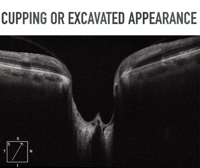
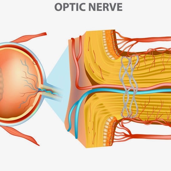
      Stereoscopic ophthalmoscopy demonstrates an ONH with “cupping” or an “excavated appearance” from RGC axonal death near the peripapillary retina, disc, or anterior lamina cribrosa. This “cupping” or “excavated appearance” is an uncommon ONH finding considered highly pathognomonic for glaucoma. Clinical examination may reveal focal optic nerve rim thinning, vertically elongated cup-to-disc ratio, vessel bayoneting, peripapillary atrophy, or optic disc hemorrhages. Perimetry displays VF defects related to nerve fiber bundle damage such as: nasal step, arcuate scotoma, hemifield defect, or generalized defects commonly seen in advanced glaucoma.

      SYMPTOMS OF INITIAL AND ADVANCED GLAUCOMA

      Initial Glaucoma: One of the challenges of glaucoma diagnosis and management is the initially asymptomatic nature of the condition. Patients usually experience no symptoms until advanced stages. Conditions, like glaucoma, in which symptoms do not match structural severity are known as having structural-functional dissociation. At least 30-40% of the optic nerve tissue must be damaged before such structural abnormality will lead to functional symptoms of vision loss. Absence of ocular symptoms initially (especially when progressing slowly) seem to be masked by brain plasticity, as the brain compensates for diminished visual signaling. Another proposed explanation is the phenomenon of “habituation” to slow eyesight deterioration developing and progressing over a long period of time. This is likely why those afflicted by glaucoma rarely complain of vision loss nor consider such eyesight deterioration as “visually threatening symptoms”.

      Nevertheless, some vision changes or signs of vision deterioration may be evident in this stage, albeit rare. Unrealized symptoms of glaucoma in the stage include increased light sensitivity, reduced nighttime vision, and slight to moderate “cloudiness” or “foggy” vision.

      Advanced Glaucoma: In advanced stages of GON, patients undeniably begin experiencing symptoms of constricted peripheral vision oftentimes resulting in difficulty navigating around rooms and objects. Patients may mention bumping into walls and objects while walking. At this time, specific symptoms emerge including “hazy” or “foggy” vision, intense glare, and severely reduced vision in low light environments. At an initial ophthalmological examination, such symptoms immediately place glaucoma on the differential diagnosis list. Tragically, end stage glaucoma also results in diminished central vision or even total blindness. 

      Summary

      Glaucoma is normally diagnosed at a routine eye examination or incidentally discovered during an eye examination for a specific reason (such as an annual diabetic eye examination). The patient is almost always asymptomatic at the time of diagnosis. Glaucoma being largely asymptomatic sometimes results in patients struggling to remain compliant with medicinal treatments, as no symptoms remind patients of potential impending vision loss.

      Ocular Hypertension:
      High Pressure Without Glaucoma

      Glaucoma Eye Pressure Check Lowering Ocular Hypertension Restore Vision

      Approximately 3-6 million Americans and 4-7% of people above the age of 40 years old have elevated IOP without evidence of glaucomatous optic nerve damage. Normal IOP ranges from 10-21 mmHg with a mean of 16 mmHg. Therefore an IOP of greater than 21 classifies an eye as being ocular hypertensive. This increased pressure may result from decreased aqueous drainage, increased aqueous production, or could be that eye’s normal, as IOP is measured on a bell curve. Patients with ocular hypertension (OHTN) do not experience any pressure sensation or blurred vision. OHTN is not accompanied by cupping of the optic disc, VF defects, thinning of the RNFL, or RGC loss, as this would denote glaucoma, not OHTN. Therefore, patients with high IOP in the absence of glaucomatous damage are referred to as having OHTN or being glaucoma suspects

      As no established standard of care existed for OHTN patients, the Ocular Hypertension Treatment Study (OHTS) was designed to evaluate the efficacy and benefits of initiating IOP lowering treatment to delay or prevent the onset of glaucoma. OHTS found that 5 years later the cumulative probability of glaucoma development for patients taking IOP lowering medication was only 4.4% and 9.5% in the untreated observation group. The OHTS results do not suggest that all patients with elevated IOP should be treated with ocular hypotensive medication and the researchers admit adverse effects of prolonged and indefinite medication use may exist, especially regarding financial and quality of life burden. Importantly, this study also identified risk factors for glaucoma, which were mentioned previously.

      Fedorov Restore Vision Clinic Berlin Germany Anton Fedorov

      Notably, ONLY 9.5% of untreated OHTN patients developed glaucoma within 5 years. OHTS identified pertinent risk factors increasing the likelihood of OHTN patients eventually developing glaucoma. From the OHTS results, and similar studies, one can gather that ocular hypertension alone is not a sufficient or necessary factor for glaucoma development. Additionally, untreated OHTN patients have less than a 10% chance of developing glaucoma 5 years later. Said differently, over 90% of untreated OHTN patients will not proceed to develop glaucoma within 5 years. Such findings clearly demonstrate that while high IOP may raise the risk of glaucoma development and progression, high IOP alone does not warrant treatment with ocular hypotensive medication. Furthermore, 4.4% of the OHTN patients successfully treated with IOP lowering medication STILL developed glaucoma within 5 years. Moreover, the majority of major glaucoma studies (Advanced Glaucoma Intervention Study, Collaborative Normal Tension Glaucoma Study, Collaborative Initial Glaucoma Treatment Study, Early Manifest Glaucoma Trial) all demonstrate glaucoma progression despite significantly lowering IOP. These well-established outcomes indicate that glaucoma is far more complicated and multifaceted than elevated or fluctuating IOP alone.

      Likewise, new research indicates there is no compelling evidence that postoperative transient IOP spikes in healthy eyes threaten optic nerve health and warrant treatment. The cessation of unnecessary IOP lowering treatments lessens clinician workload, decreases cost of medication for patients, reduces patient visits, and minimizes potential clinical and subclinical medication side effects.


      THE ROLE OF THE VASCULAR SYSTEM IN GLAUCOMA INITIATION AND PROGRESSION

      Eye care practitioners evaluate glaucoma progression based on both structural and functional metrics. Structural measurements are obtained using optical coherence tomography (OCT) technology calculating the thickness of the axons surrounding the ONH. Guided progression analysis (GPA) software analyzes data over time to quantify the rate of change while plotting these results on a graph. This graph depicts the current rate of progression and even predicts further progression over time. Functional progression is determined by field of vision loss estimated through automated perimetry and compared with previous values. These data sets indicate if progression is “possible”, “likely”, or “confirmed”.

      Once progression is confirmed - a “target IOP” is determined using current IOP, percentage of desired IOP reduction, and degree of structural and functional loss. As current medicinal and surgical glaucoma treatments currently only focus on IOP lowering, a “target IOP” is formulated for each patient. Although studies validate that lowering IOP lessens the rate of glaucoma progression, these studies also confirmed that glaucoma frequently progresses despite achieving this “target IOP”. Such evidence implies factors other than IOP contribute to glaucoma progression.


      A VASCULOPATHIC VIEW OF GLAUCOMA

      ischemic optic neuropathy ischemia anterior portion optic nerve Sleep Apnea

      While elevated IOP is an established risk factor for progression, perhaps a more significant risk factor is low ocular blood flow or ocular perfusion pressure (OPP). OPP is defined as the difference between arterial blood pressure and IOP. Therefore, both a high IOP and a low BP will lessen OPP potentially instigating ischemic damage to the retina and optic nerve. Research indicates that patients with low OPP have a considerably higher risk of glaucoma progression compared to patients with high OPP. Also, progression is associated with fluctuations in OPP from inconsistent BP or IOP. Nocturnal BP is normally lower due to diurnal patterns, however studies confirm progressive glaucomatous VF loss occurs in cases of exaggerated nocturnal BP drop.

      The Thessaloniki Eye Study demonstrated increased cupping and decreased ONH rim area in non-glaucomatous patients taking anti-hypertensive medication whose diastolic BP fell below 90 mmHg overnight. Similarly, a BP drop of 10% or more in glaucoma patients with well-managed hypertension was connected with structural ONH damage and progressive VF loss. Also, the magnitude of OPP fluctuations is correlated with glaucoma severity in NTG patients.

      New vascular imaging methods demonstrate reduced ocular blood flow (OBF) in glaucoma patients, especially in cases of progression despite normal IOP. Moreover, glaucoma progression can even be forecasted by diminished OBF rates. Fluctuations of ocular blood flow are particularly damaging and may be a more significant measurement for progression than baseline OBF measurements.

      HOW THE EYE AND BRAIN COMPENSATE FOR REDUCED OCULAR BLOOD FLOW

      The intrinsic capability of vascular autoregulation serves to continuously provide oxygen and nutrients despite any metabolic or pressure irregularities. The importance of autoregulation lies in the ability to maintain constant ocular blood flow despite (OBF) inconsistent ocular perfusion pressure (OPP). The autoregulatory response alters OBF in response to variations in OPP. Though elevated IOP results in decreased OPP, normal autoregulation enables microvascular consistency thus preventing ischemic damage. However, the ONH becomes vulnerable to OPP fluctuations if autoregulation is compromised or its regulatory range is surpassed. Such autoregulation impairment is one potential vasculopathic mechanism for the risk and progression of GON despite relatively normal IOP and BP fluctuations. Though OPP may decrease as a result of high IOP or low BP, research has not clearly demonstrated which, if either, is more injurious to the ONH. However, research suggests this autoregulation system better regulates low OPP induced by high IOP than that of low choroidal BP, suggesting that low blood pressure may play a more significant role than high IOP in GON. Also, in glaucoma patients with both normal and elevated IOP, studies reveal lower BP and greater nocturnal BP variation.

      While the ONH blood supply through the parapapillary choroid can normally autoregulate to maintain a constant OPP, significant changes in BP or IOP may exceed the autoregulation ability. When either the range of autoregulation is exceeded or autoregulation function is impaired, the ONH is at risk for ischemic injury. Studies also indicate dysregulation or even non-regulation of ocular vasculature in glaucoma patients. Such dysregulation or non-regulation poses a substantial threat to the ONH with normal BP and IOP fluctuations, let alone abnormal fluctuations. Moreover, loss of autoregulatory function may further predispose an ONH to GON when subjected to high IOP or low BP.

      Some researchers believe systemic primary vascular dysregulation (PVD) is the source of ocular vascular dysfunction triggering GON. While some optic neuropathies are caused by hypoxia, the fluctuations in OBF triggering oxidative stress may contribute to glaucoma risk and progression. PVD commonly results in stiff and irregular retinal vessels, increased retinal venous pressure, impaired neuro-coupling, and decreased autoregulation capacity, all of which increase glaucoma risk and progression. 

      Evidence now exists that when vascular autoregulation is compromised, even normal BP and IOP fluctuation may lead to inconsistent OPP and subsequent ischemic ONH damage.  For this reason, primary vascular dysregulation is a substantial risk factor for GON as it is associated with impaired autoregulation, low nocturnal BP, and increased retinal venous pressure – each of which can cause GON independently. It is reasonable to assume that reduced OBF from dysfunctional autoregulation may play a significant role in GON risk and progression. Furthermore, research findings suggest proper autoregulatory responses are altered or missing with glaucoma. These daily and repetitive ischemic events suggest a potential vascular mechanism in glaucoma, especially NTG. Despite this, vascular etiology in glaucoma is rarely considered amongst eye care professionals, even in NTG eyes with vasculopathic risk factors. 

      This evidence strongly suggests that elevated IOP alone does not cause GON, but rather diminished ocular blood flow, which is exacerbated by elevated IOP and reduced BP. While a vascular autoregulation system exists, PVD may limit or abolish this regulation capability leading to reduced OBF and subsequent ONH ischemic damage. Similarly, autoregulation capabilities may be exceeded in cases of elevated IOP, low BP, or OPP fluctuation. For these reasons, glaucoma progression is not resultant from high IOP or low BP alone, but in conjunction with decreased OPP resulting in ONH ischemia, axonal loss, and RGC death. This may explain why cardiovascular disease, hypertension, and diabetes are risk factors for glaucoma progression. Such evidence is biologically plausible and suggestive of a vasculopathic mechanism for progression.

      CEREBROSPINAL FLUID PRESSURE AND GLAUCOMA

      Idiopathic Intracranial Hypertension Intracranil Pressure Cerebrospinal fluid Optic Nerve Treatment Restore Vision Clinic

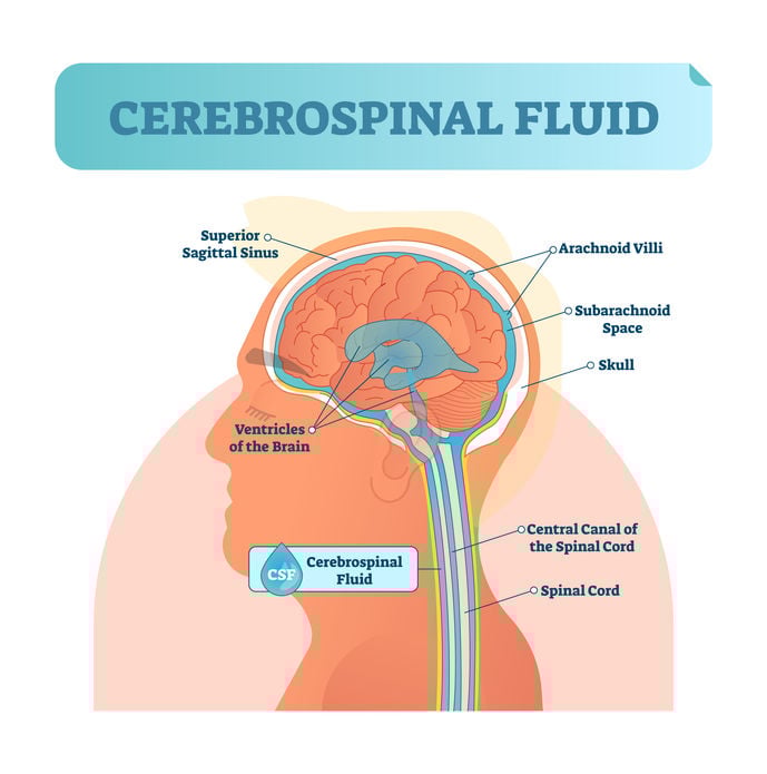
      As research continues to support the concept of multiple pressure types having roles in GON, new findings suggest a previously overlooked intracranial pressure that may contribute to GON. Interestingly, initial studies in cats indicate an association between low cerebrospinal fluid (CSF) pressure and glaucoma development. Relatedly, human subjects with NTG have significantly lower CSF pressure compared to normal subjects. Similar research reveals that NTG subjects also have appreciably narrower orbital CSF space, which is associated with lower CSF pressure, when compared to high IOP glaucoma subjects. Given that glaucoma still develops and progresses despite normal IOP, it is plausible that NTG development and progression may partially arise from low orbital CSF pressure related to narrower orbital CSF space. Though research is ongoing, these initial results suggest that pressure behind the eyes can be as significant as pressure inside the eyes, especially in NTG patients.

      Glaucoma Brain degeneration Restore Vision Clinic (1)

      IS GLAUCOMA AN EYE DISEASE ONLY? BRAIN IN FOCUS

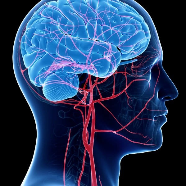
      As previously mentioned, though elevated IOP is certainly a risk factor for glaucoma progression, vision deterioration often still occurs despite successfully lowering IOP using medicinal and surgical treatments. Such instances suggest an IOP independent mechanism triggering this progression. Neuroimaging technology can potentially shed light on another side of glaucoma. For decades, glaucoma has been classified as a neurodegenerative disease by many researchers and clinicians. Similarities exist between neurodegenerative conditions, specifically Parkinson’s and Alzheimer’s diseases, and glaucoma regarding loss of neurons, transsynaptic degeneration, and cell death. Recent neuroimaging findings demonstrate that glaucoma patients have focal brain damage in other brain areas besides the retina and optic nerve. Brain tomography reveals structural changes of the lateral geniculate nucleus (LGN), optic radiations, and visual cortex associated with glaucoma.

      Remarkably, structural changes have even been observed in non-visual areas of the brain. New fascinating evidence is emerging showing that glaucoma patients undergo atrophy of the amygdala potentially resulting in emotional and memory related complications. Such abnormalities of the amygdala may explain why those afflicted with glaucoma often experience mood instability, increased anxiety, heightened fear, and feelings of anger.

      Given these new discoveries, glaucoma may be either a primary neurodegenerative disease or a primary optic neuropathy with secondary CNS damage. Provided that VF defects in glaucoma are related more to nerve bundle fiber patterns rather than LGN injuries, glaucoma is likely a primary optic neuropathy with secondary neurodegenerative involvement. Nonetheless, glaucoma is not a disease limited to the eye, but is now also recognized as a brain disease.

      Given these new discoveries, glaucoma may be either a primary neurodegenerative disease or a primary optic neuropathy with secondary CNS damage. Provided that VF defects in glaucoma are related more to nerve bundle fiber patterns rather than LGN injuries, glaucoma is likely a primary optic neuropathy with secondary neurodegenerative involvement. Nonetheless, glaucoma is not a disease limited to the eye, but is now also recognized as a brain disease.

      MANAGEMENT OF GLAUCOMA
      AND THE VASCULAR SYSTEM

      Currently all glaucoma treatment models are centered on lowering IOP, which can potentially lead to the misinformation of glaucoma being caused by high IOP only. Topical medications, oral medications, laser surgeries, and implants all attempt to limit glaucoma progression through IOP reduction. It's well known that some patients continue to progress despite a low or normal IOP which further suggests an IOP-independent mechanism at play. 

      VASCULAR SYSTEM MANAGEMENT

      Research suggests vascular dysfunction being a primary component in glaucoma, rather than occurring secondarily. In cases of systemic vascular disease blood vessels are stiffer, more irregular, demonstrate reduced neuro-coupling, and have diminished autoregulation function. Such abnormalities are not isolated systemically, but affect the eyes as well. Due to the vascular nature of glaucoma, it is reasonable to conclude that what is healthy for the vascular system is healthy for ocular blood supply. Alleviating risks factors of vascular diseases such as hypertension, diabetes, and cardiovascular disease likely similarly lessen glaucoma risk and progression. Eye care providers should also communicate better with primary care physicians, regarding proper use of anti-hypertensive medications as this may markedly affect OPP thus provoking glaucomatous damage.

      CONCLUSION

      Glaucoma is perhaps more multifaceted and less understood than all other eye conditions given the ocular, vascular, and neurological involvement. Glaucoma is a collection of progressive optic neuropathies with specific ONH appearance, RGC axonal loss, and VF defects. Much confusion stems from glaucoma being neurodegenerative, having vascular etiology, yet only IOP being commonly discussed and treated. While IOP is certainly a risk factor, progression of glaucoma is likely more so related to ischemia from diminished ocular perfusion pressure given that progression may occur with any IOP. Glaucoma terminology, thought processes, and treatments are obsolete resulting in inadequate patient education and poor outcomes. In the future, glaucoma mechanisms will be better understood and improved treatments will be developed limiting glaucoma development and progression.


      Blog prepared in cooperation with Kaleb Abbott, O.D., M.S.


      Driving Low Vision Fedorov Clinic Restoration Therapy  Berlin GermanyNote: The information given in this blog are the opinions of the authors and for reader familiarization purposes only. This blog is not intended as a substitute for professional medical advice. Also, the information provided does not replace or abolish any official or legal terms for glaucoma diagnosis, treatment, and management. Authors are not liable for any undesirable consequences or effects related to the information provided in the blog.

      REFERENCES

      1.     Vrabec JP, Levin LA. The neurobiology of cell death in glaucoma. Eye (Lond). 2007;21 Suppl 1:S11-4.
      2.     Lee KM, Kim JM, Lee EJ, Kim TW. Anterior Optic Nerve Head Perfusion is Dependent on Adjacent Parapapillary Choroidal perfusion. Sci Rep. 2019;9(1):10999.
      3.     Leske MC. Ocular perfusion pressure and glaucoma: clinical trial and epidemiologic findings. Curr Opin Ophthalmol. 2009;20(2):73-8.
      4.     Kass MA, Heuer DK, Higginbotham EJ, et al. The Ocular Hypertension Treatment Study: a randomized trial determines that topical ocular hypotensive medication delays or prevents the onset of primary open-angle glaucoma. Arch Ophthalmol. 2002;120(6):701-13.
      5.     Jonas JB, Wang N. Cerebrospinal fluid pressure and glaucoma. J Ophthalmic Vis Res. 2013;8(3):257-63.
      6.     Wang J, Li T, Sabel BA, et al. Structural brain alterations in primary open angle glaucoma: a 3T MRI study. Sci Rep. 2016;6:18969.
      7.     Flammer J, Konieczka K, Flammer AJ. The primary vascular dysregulation syndrome: implications for eye diseases. EPMA J. 2013;4(1):14.
      8. Glaucoma Structural and Functional Progression in American and Korean Cohorts Ophthalmology. 2016 April ; 123(4): 783–788. doi:10.1016/j.ophtha.2015.12.010.

      Find out if you are a good candidate

      Get in touch today