STRUCTURE, FUNCTION, DAMAGE / DISORDERS, AND TREATMENT

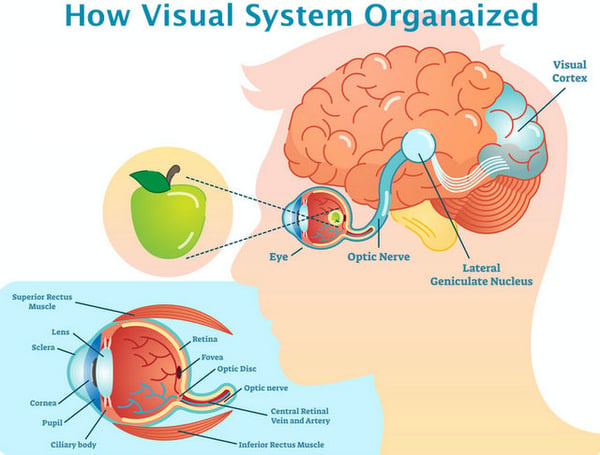
      What is this nerve and why is it so essential to our sight? To answer this question we first have to understand what actually takes place in the physical act of seeing. Our visual system is comprised of three major parts: the eye, the visual cortex (located in the brain), and the pathway that connects them. Visual System Structure Field of Vision Fedorov Restore Vision Clinic
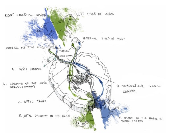
Within the eye is the retina, the cells of which are responsible for perceiving, processing, and relaying information to the brain, where the decoding of the received signals takes place and our actual visual experience occurs. The eye and brain are intricate, complexly structured organs, and their function in the visual process can’t be performed correctly if they’re not properly connected to each other. This is why the optic nerve, acting as a type of bridge between the two, plays such a critical role in how we see the world.

      The optic nerve (occasionally referred to as cranial nerve II or CN II) is a structure consisting of millions of fibers and is responsible for everything from the dilation and contraction of the pupil (the light reflex) to how words and images are viewed up close or at a distance. Essentially, all visual signals are transmitted from the eye to the brain via this pathway, and as a result any injury to this nerve structure can cause vision loss, with the level of impairment depending on the severity and location of the damage. In turn, such damage can lead to drastic consequences for our lives and overall wellness.

      Retina Optic Nerve Visual Cortex Fedorov Restore Vision Clinic

      Every day, thousands of people suffer from vision loss caused by optic nerve damage, and many individuals complain not only about their limited vision, but also limitations within the healthcare system itself. In fact, many visually impaired patients and their family members find that their healthcare providers do not treat their condition at all, or that the type of treatment offered doesn’t appropriately address their particular problem. These struggles—along with the difficulty of finding reliable information regarding various conditions and different treatments available—can cause considerable stress and doubt.

      However, once a healthcare provider who attends to those with optic nerve damage is found, and relevant, practical information is obtained, patients can more accurately determine the most effective treatment method available, and whether or not their vision loss can be stabilized or partially reversed. This page is designed to provide such information and address the issues mentioned above, offering you an in-depth look at the optic nerve. It will guide you toward a greater understanding not only of what it is and how it works, but also what types of damage and disorders can occur and the various ways they can be treated.


      STRUCTURE OF THE OPTIC NERVES

      HOW OPTIC NERVES GROW AND DEVELOP

      The primary development of the human eye occurs between weeks three and 10 of the human gestational period. During the seventh week, the optic nerves—dual cylindrical structures that extend (one each) from the rear of the eyes—are derived from structures known as optic stalks in earlier stages of development. The evolution of the nerves continues through gestation and is sustained through the early years and into adolescence. During this term of development, very specific features and pathways are created which allow information received from the eye to pass successfully to the brain. An understanding of the structure, composition and visual pathway of the optic nerve (see figures above) is crucial for achieving a sense of its function and importance to the operation of sight.

      STRUCTURE

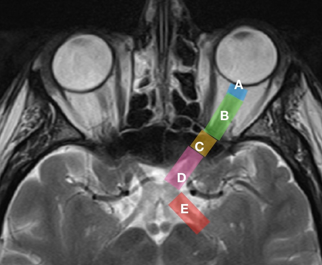
      The optic nerve varies in length from person to person—and even differs between the two eyes of a single individual—but generally measures 35-55 mm. The most efficient way to describe the structure of the nerve is by dividing it into four main sections: (A) optic nerve head (or intraocular part), (B) intraorbital part, (C) intracanalicular part, and (D) intracranial part.

      Optic nerve structure parts Fedorov Restore Vision Clinic

      OPTIC NERVE HEAD (A)

      The optic nerve head is a very unique part of the optic nerve as it marks a pivotal point of vascular, geometric and tonometric transformations.  The optic nerve head—which may also be referred to as the optic disc or, more rarely, the “papilla”—is approximately 1mm in length and 1.5mm in diameter. The diameter and arrangement of the optic disc and the corresponding physiological cup depends upon the variable shape, size, and orientation of the opening into the Bruch’s membrane and the conically shaped chorioscleral canal. 

      The optic nerve head is a distinctive part of the nerve as a whole, and can itself be subdivided into three aspects: the surface nerve fiber layer, the prelaminar region and the lamina cribrosa region.

      The surface nerve fiber layer—the most anterior (closest to the front) layer of the optic disc—incorporates the densely wound optic nerve fibers stemming from retinal ganglion cells across the whole retina as they merge and angle toward the optic nerve. This layer consists of a high volume of blood vessels, including sizable retinal vessels, venous channels and a tightly packed complex of capillaries.

      The prelaminar region lies directly behind the surface nerve fiber layer, and is generally called the anterior part of the lamina cribrosa, although it can also be referred to as the glial or choroidal part. This region is one of the most essential areas of the optic nerve. It is composed primarily of glial tissue, a type of binding or connective tissue found throughout various parts of the nervous system. Glial fibers run perpendicularly to the nerve fiber masses and are significantly thinner than the coarser connective tissue fibers. This region also hosts a high number of capillaries surrounded by a membrane, which serve to aid and protect the nerve fibers as well as provide them with nourishment.

      Where the prelaminar region of the optic nerve head is the anterior part of the lamina cribrosa, the lamina cribrosa region is known as the posterior part of the lamina cribrosa (or the scleral part). 

      The lamina cribrosa is a mesh-like structure composed of dense collections of collagen fibers as well as glial sheets. Similar to the prelaminar region, the nerve fiber groupings are protected by a glial membrane composed of glial tissue. The lamina cribrosa is central to regulating pressure levels between the interior of eye and the tissues that surround it.

       INTRAORBITAL PART (B)

      This part of the optic nerve, roughly 25mm in length, runs from the back of the eyeball to the opening of the optic canal. In this region, the optic nerve’s diameter is approximately twice that of the intraocular part, making it roughly 3-4mm wide.  This is due to the myelination of the nerve fibers in this region and onward along the nerve’s length.

      EYE OPTIC NERVE CHIASMA FEDOROV RESTORE VISION CLINIC

       The myelin sheath is an extended and modified membrane wrapped around the optic nerve in a spiral fashion and aimed to be an electrical insulator. The function of myelin is to increase the speed of nerve conduction and neural circuits, and myelination serves as a source of plasticity in neural circuits. 

      This portion of the optic nerve is also surrounded by the meningeal sheath, which is made up of the arachnoid mater, pia mater and dura mater (the three meningeal layers), as well as cerebrospinal fluid. The dura mater and arachnoid mater are loosely joined near the back of the eyeball. Here there is greater subarachnoid space between the nerve and the sheath, creating a bulbous area behind the eye;  the subarachnoid space between the nerve and the sheathnarrows as the nerve approaches the optic canal. 

      This part of the nerve contains roughly textured connective-tissue septa (septa being the plural of septum, a partition between chambers). These septa contain blood vessels, branch out in all directions, and are all connected to each other. It should also be noted that this part of the nerve rests in close proximity to the ophthalmic artery.

      INTRACANALICULAR PART (C)

      This part of the nerve lies within the bony optic canal and has a length ranging from 4-10mm. As with the intraorbital part, this portion of the nerve is also enclosed by the meningeal sheath. In fact, this part is virtually identical in its basic structure to the intraorbital section of the nerve discussed above. 

      INTRACRANIAL PART (D)

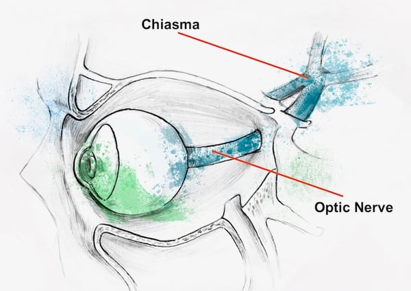
      Finally, this section of the nerve, approximately 10mm long, extends from the rear opening of the optic canal, continues on above the diaphragma sellae and the cavernous sinus. At this point the optic nerve from one eye joins with that of the other eye, creating the optic chiasm.

      OPTIC NERVE: ANTERIOR

      The optic nerves are composed from axons (fibers) of retinal ganglion cells which gather together forming a trunk at the optic nerve, which then passes through the optic canal, within the cranial bones, and continues on to an intersection called the optic chiasm. The optic chiasm is a cross-like structure formed by the interweaving of nerve fibers.  From this junction, the nerve fibers—now called “optic tracts”—extend to the lateral geniculate nucleus (or LGN) located in the thalamus. The whole of the thalamus is an essential relay center for sensory information received from various receptors, including the retina. It executes a primary analysis of incoming signals before their arrival at the occipital lobe of the brain, where the visual cortex is located. The LGN neuron’s fibers compose the next level of our visual pathway called optic radiation, which are responsible for conveying sensory information to the brain for further processing. Within the brain, the visual cortex then decodes signals from the retina and translates them into the real visual images we experience. This ultimately results in what we call vision.

      OPTIC NERVE: POSTERIOR (E)

      As you can see, even this general overview of the visual pathway can be somewhat difficult to understand. However, it can be simplified further by taking a closer look at the origins of this process: the cornea, lens, and particularly the retina and its cells.

      The physical operation of sight is initiated when light reflected from objects passes through the cornea (the clear exterior part of the eye), and on through the lens. The lens’s primary purpose is to focus and direct that light onto the retina at the back of the eye, where chemical and electrical impulses (sensory information) are generated and passed on through the visual pathway to the brain. The retina’s connection to the optic nerve is essential to understanding the visual process and the nerve itself.

      The retina is composed of several layers, including the layer of photoreceptor cells (two varieties: rods and cones), bipolar cells and retinal ganglion cells. Rods are responsible for processing black and white vision (including dim or low light), as well as our peripheral (or side) field of vision, which comes from signals captured by the rods. Cones are necessary for perceiving color and also play a role in our central vision, the type of vision being used while you read this text, for example. Before the information sent via the rods and cones can continue on its journey, it must be processed further by neurons, which produce high amplitude electric impulses (or “action potentials”). 

      These neurons are called Retinal Ganglion Cells, or RGCs, of which there are roughly one million in the retina. While these cells are variable in size and other physical and functional aspects, they share one key characteristic: each of them has a lengthy axon—a fibrous, thread-like part of a nerve cell—that physically extends and carries impulses all the way to the brain. RGCs are connected to corresponding neurons of the visual cortex, which, again, provides an analysis of the information obtained from the outside environment. In other words, the fibers from the RGCs are literally the building fibres of which the optic nerve is composed.

      SUMMARY

      Clearly, the optic nerve—its parts, composition and pathway—is amazingly intricate and highly complex. Its basic function, however, is relatively straightforward. It is an essential part of the central nervous system that serves to transmit it to the brain where that data is ultimately processed. In other words, structures— like our retina, optic nerve, higher visual pathways and subcortical visual centers—are essential and integral parts of a complex visual system in which data is gathered, transmitted and partially decoded. Keeping this core function in mind will help patients and information seekers better understand the centrality of the optic nerve to their overall health and wellness.

      Diseases of the Optic Nerve

      Both diseases and pathological processes of the optic nerve can induce optic nerve damage, injury and alteration, each of which is characterized by structural abnormalities typical of and particular to the origin of the damage, and can result in the partial or complete loss of optic nerve function and, ultimately, to vision loss . The severity of vision loss generally corresponds directly to the extent of structural damage.

      During the acute stage, both structural changes of the optic nerve and vision loss can be reversed. However, if no symptomatic treatment is applied during this period, then optic neuropathy enters the chronic stage when structural and functional alterations can no longer return to normal conditions. This, in turn, means that early acute damage of the optic nerve has passed into various stages of  Optic Nerve Atrophy/Degeneration.

      The classification of optic nerve diseases and pathological processes remains incomplete, and more common definitions of optic neuropathy are based on various aetiologies (or cause/(s)) of damage and different types of optic nerve atrophy (anterograde, retrograde and trans-synaptic degeneration).

      The origins (aetiology) of optic nerve damage may be divided into two groups, depending on the location of the given pathological process: within or in close proximity to the eyeball (ocular), or remote from the eye, nearer to the brain (cerebral). The management of both medical conditions tends to be different. Ocular medical conditions include blood supply failures, local infections and local events such as optic nerve compression. Several hereditary diseases and developmental anomalies affect optic nerve function at the level of the eyeball. Cerebral causes are the most remote from the eye itself but damage the optic nerve along its pathway from the  eye to the subcortical brain’s visual center (located in the thalamus). In these cases, typical causes of damage include elevated intracranial pressure, local pathological processes along optic pathways, common pathological factors such as inflammation, autoimmune processes and various intoxications. Most of these cause secondary damage of the optic nerve.

      OPTIC NEUROPATHY CAUSED BY PATHOLOGICAL PROCESSES IN OR NEAR THE EYE.

      Idiopathic Intracranial Hypertension Neuroimaging MRI CT Restore Vision Clinic

      Optic nerve tumors

      The most commonly known tumors are optic nerve gliomas, primary sheath meningioma, melanocytoma, malignant astrocytoma, astrocytic hamartoma, lymphoreticular tumors and metastatic tumors. Optic nerve tumors can be divided into primary tumors—which are tumors of the nerve—and tumors arising from the sheath. The most common tumor of the optic nerve is the optic nerve glioma, which are usually benign. However, some tumors such as gangliogliomas, medulloepitheliomas, haemangioblastomas and haemangiopericytomas are malignant gliomas. Meningioma is the most typical tumor of the optic nerve sheath. Primary sheath meningiomas arise from intraorbital or intracanalicular portions of the optic nerve. Secondary meningiomas are from intracranial sources. Most tumors cause gradual visual loss which progresses slowly and is clinically associated with further evidence of an anterior or posterior optic neuropathy, as well as compressive metastases and tuberculomas.

      Ophthalmic Associations Obstructive Sleep Apnea Glaucoma

      Glaucoma

      Glaucoma a leading cause of blindness—is a group of eye diseases that progressively and, in many cases, silently damages the optic nerve causing gradual and permanent vision loss. Generally, glaucoma is associated with increased fluid pressure within the eye, or intraocular pressure (IOP). At present, further damage to the optic nerve—or glaucomatous optic neuropathy—is avoided by lowering IOP, and the effectiveness of glaucoma treatment and management is measured by how well IOP is controlled. Reducing eye-fluid pressure in the eye through medications (eye drops) or surgery is a standard approach for treating glaucoma. But even with IOP lowered and stabilized, vision loss is not definitively prevented.

      In such cases, our treatment can be used as an attempt to produce changes of sight. Taking into account that even in cases of severe vision deterioration or in the absence of formal vision, 10% to 15% of our patients have an ability to achieve some positive dynamics, we cannot completely exclude the recommendation of conducting a course of stimulation.

      retinitis-pigmentosa-treatment--our-treatment--therapeutic-electrical-stimulations-thumb-2

      Anterior ischemic optic neuropathy leading to ischemia/infarction of the nerve tissue.


      Depending on the segment of the optic nerve affected, ischemic optic neuropathies (IONs) are divided into anterior and posterior categories. Anterior IONs (AION) are subdivided into nonarteritic and arteritic etiologies. Arteritic anterior ischemic optic neuropathy (AAION) is caused by inflammation of arteries and requires immediate therapy to prevent blindness. The most common disorder associated with arteritic AION is giant cell arteritis (GCA). Non-arteritic ischemic optic neuropathy (NAION) is more common than arteritic AION, accounting for up to 95% of ischemic optic neuropathies. This condition is the result of the occlusion (or blockage) of small blood vessels supplying blood to the optic nerve head.

      Idiopathic Intracranial Hypertension Neuroimaging MRI CT Restore Vision Clinic Berlin

      Optic nerve damage due to ocular (eye) trauma.

      Traumatic optic neuropathy are subdivided into direct and indirect forms. Direct injury is the result of penetrating eye trauma, seen frequently with orbital fractures. Several varieties of direct optic nerve injury may be revealed as optic nerve avulsion, transection, optic nerve sheath haemorrhage, orbital haemorrhage and orbital emphysema. Avulsion usually follows severe orbital trauma with severe and immediate vision loss. Optic nerve transection occurs as the result of facial trauma or orbital fracture.  Optic nerve sheath haemorrhaging causes potentially reversible visual loss, as opposed to the aforementioned conditions. Bleeding in the optic nerve sheath can be drained via a sheath fenestration leading to functional recovery (and an improvement in vision). Orbital haemorrhaging typically injures the optic nerve due to raised pressure within the orbit.

      Leber Optic Atrophy

      Hereditary degeneration of the optic nerve

      Hereditary optic neuropathies comprise a group of disorders which includes Leber Hereditary Optic Neuropathy (LHON) and Autosomal Dominant Optic Atrophy (DOA), also known as Kjer disease. Major lesions can be seen in papillomacular bundles which lead to the loss of central vision. This results in massive retinal ganglion cells (RGC) loss, seen clearly in the central region of the retina.  More devastating, however, is that in most cases hereditary optic neuropathies are progressive. The pattern of transmission of the genetic deficit is employed for the classification of inherited optic neuropathies. They are autosomal dominant, autosomal recessive and mitochondrial. Often times, optic nerve dysfunction is simply a manifestation of more common diseases, which include various neurologic and systemic manifestations that cause multi-system degenerations.

      Inflammation of the optic nerve (Optic Neuritis, Neuromyelitis Optica and Neuromyelitis Optica Spectrum Disorders (NMOSD)

      Retinitis Pigmentosa Restoring Vision Fedorov clinic restore vision

      Optic neuritis is an inflammatory condition affecting the optic nerve. It is idiopathic (or of an unknown cause) in most cases, but carries a strong association with multiple sclerosis (MS),  where the demyelinating process is the main cause for developed optic nerve damage. In rare cases, other etiologies like infectious, inflammatory, and other pathological immunological responses play a role in this medical condition. Based on recommendations of North American Neuro-Ophthalmology Society (2012), optic neuritis is a clinical diagnosis related either to inflammatory demyelination of the axons connected with diagnosed multiple sclerosis (MS), or to idiopathic conditions, which typically occurred at age 20-50, of which approximately 75% are female. 

      At the time of presentation, acute visual loss is typical and is generally monocular and accompanied by peri-orbital pain. If there are no such signs present, atypical optic neuritis must be considered, which presents itself in the following forms: neuromyelitis optica, neuroretinitis and chronic recurrent immune optic neuropathy. In 1-3% of all forms of optic neuritis, neuromyelitis optica (or Devic Disease) is diagnosed, which is by nature an autoimmune disease of the central nervous system and is characterized by inflammatory demyelinating lesions in the spinal cord and optic nerve.

      Neuromyelitis Optica (NMO) must be distinguished from multiple sclerosis as it is a  completely different type of autoimmune damage leading to the appearance of specific antibodies against the astrocytic water channel aquaporin-4 (AQP4). Simultaneously with diagnosed  bilateral optic neuritis, the spinal cord can be damaged, with such damage known as longitudinally extensive transverse myelitis. Frequently there is a gap oftwo to four years between the onset of optic neuritis and the onset of transverse myelitis. In the last few decades, the definition of “Neuromyelitis Optica (NMO)“ has been extended to “NMO spectrum disorders” (NMOSD) when there includes an involvement of almost any CNS region, as well as cases in which restricted involvement of a single region can be seen.

      Developmental anomalies

      (optic nerve hypoplasia, colobomas, retarded myelination, congenital swelling of the optic disc (pseudo papilloedema).
      Optic Nerve Hypoplasia Fedorov Restoration Therapy electrical stimulation Restore Vision Clinic Berlin

      Optic nerve hypoplasia (ONH) is characterised  by an abnormally small optic nerve head frequently seen together with head cupping. Loss of vision occurs because the number of axons in the optic nerve are limited due to apoptosis during the development stages of the visual system. ONH is a unilateral or bilateral non-progressive underdevelopment of the optic nerve, and is considered to be a non-local syndrome rather than a more diffuse condition. It can be divided into three clinical subtypes: a) Optic Nerve Hypoplasia Simplex; b) Septo-optic dysplasia (de Morsier's syndrome); and c) Septo-Optic-Pituitary Dysplasia. Septo-optic dysplasia is considered to be a combination of ONH, pituitary gland hypoplasia and midline brain abnormalities.

      Optic Nerve Hypoplasia Simplex may occur as an isolated defect or in association with other ocular abnormalities (microphthalmos, aniridia, coloboma, nystagmus and strabismus). In cases where ONH takes place simultaneously with CNS abnormalities, there are several hypotheses regarding how these pathological conditions develop. The first theory states that a malformed chiasm developed, resulting in an elongation or stretching of the optic nerves. In this case, it is generally assumed that normally developed ganglion cells have reached a malformed chiasm but cannot proceed across the midline into the optic tracts, which further leads to retrograde degeneration of the axons. The second theory postulates that, due to the abnormal development of the cerebral hemispheres and ventricular system, stretching of the optic nerves takes place and leads to their retrograde degeneration and finally to the atrophy of the ganglion cells of the retina.

      IDIOPATHIC INTRACRANIAL HYPERTENSION PAPILLEDEMA DRUSEN RESTORE VISION CLINIC

      Optic Nerve Drusen

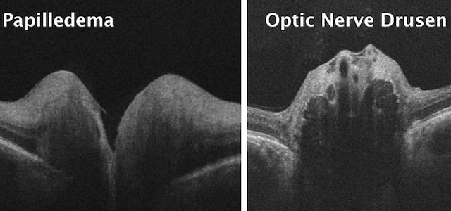
      Optic Nerve Head Drusen (ONHD) is a relatively rare hereditary anomaly with structural abnormalities caused by white calcareous deposits in the pre-laminar region at the entire disc area. In most cases, they are generally asymptomatic and bilateral. ONHD are globular, calcified bodies accumulated within the optic nerve head. It is most commonly believed that the formation of disc drusen is caused by a chronic obstruction of axoplasmic flow, which leads to the formation of deposits. There is an agreement that ONHD is an autosomal genetic determinate abnormality and typically occur in small, crowded optic discs. Many disorders have been associated with optic disc drusen. However, it seems that the only true associations are with retinitis pigmentosa (RP). If calcareous deposits mechanically compress the axons of retinal ganglion cells, then this leads to resulting defects in the field of vision. Visual field defects can progress with age, and are often detected in the second decade of life. Although ONHD are typically benign, patients with diagnosed drusen should be monitored to rule out ocular complications that can be potentially sight threatening.

      Ischaemic optic neuropathy treatment Berlin Germany

      Radiation neuropathy.

      Radiation-induced peripheral neuropathy (RIPN) is a chronic handicap which is caused by compression of the nerve by fibrosis induced by radiation. In addition there may be evidence of direct injury to optic nerves through axonal damage and demyelination or injury to blood vessels by ischaemia. Radiation therapy for intra- and extracranial tumours can affect the anterior part of the optic nerve. Ophthalmological findings are those of acute ischaemic anterior optic neuropathy with acute loss in visual acuity. However, the damage to the posterior portion of the optic nerve or chiasma is the most frequent for radiation-induced optic neuropathy, with impairment of visual function.

      In many cases, Fedorov Restoration Therapy can help improve vision – even for conditions believed to be untreatable. Our treatment approach considers the functional ability of both the vision system and the brain, and is individualized for each patient to achieve optimum results.


      Find out if you are a good candidate

      Optic Neuritis Blurry Vision Central Scotom Treatment

      Papillitis

      Papillitis is an inflammation of the optic nerve head at its exit from the eyeball, or as "intraocular optic neuritis". The inflammation can lead to an acute loss of vision. The factors that cause papillitis include infectious diseases, disseminated inflammations, intoxications, allergic-hyperergic and immunological processes. Papillitis might have the same clinical signs as papilledema. However, papillitis may be unilateral, whereas papilledema is almost always bilateral.

      Vision Loss Methanol Poisoning

      Brain tumours (especially pituitary tumours).

      Brain tumors can be malignant or benign. When benign or malignant tumors grow, they can elevate the pressure inside the skull and cause damage to brain tissue. Both benign or malignant tumours can damage the optic nerves, directly compress them, or damage them indirectly through increased pressure in the skull, ultimately causing problems with a patient’s vision. Generally brain tumors are categorized as primary or secondary. A primary brain tumor originates in the brain itself. Many primary brain tumors are benign. A secondary brain tumor, also known as a metastatic brain tumor, occurs when cancer cells spread to the brain from another organ, such as your lung or breast. Primary Brain Tumors can develop from brain cells, the membranes that surround your brain, called meninges, nerve cells or glands. In adults, the most common types of brain tumors are gliomas and meningiomas.

      Gliomas are tumors that develop from a variety of glial cells. Glial cell tumors include: astrocytic tumors such as astrocytomas, oligodendroglial tumors, glioblastomas, meningiomas, schwannomas. Other primary brain tumors include pituitary tumors (generally benign), pineal gland tumors, ependymomas, craniopharyngiomas, primary brain lymphomas, primary central nervous system (CNS) lymphomas,and  primary germ cell tumors of the brain. Secondary brain tumors make up the majority of brain cancers. They start in one part of the body and spread, or metastasize, to the brain. The following can metastasize to the brain due to lung, breast, kidney or skin cancers. Secondary brain tumors are always malignant. Benign tumors don’t spread from one part of your body to another.

      Bildschirmfoto 2018-06-15 um 13.03.35

      Autoimmune optic neuropathies

       including single isolated optic neuritis (SION), relapsing isolated optic neuritis (RION), chronic relapsing inflammatory optic neuropathy (CRION), the neuromyelitis optica spectrum disorder (NMO), multiple sclerosis associated optic neuritis (MSON) and unclassified forms (UCON). 


      Cerebral vasular pathology

      Cerebral vasular pathology is associated with diabetes, hypertension, and hypercholesterolaemia.

      Idiopathic Intracranial Hypertension Neuroimaging MRI CT Restore Vision Clinic

      Head trauma

      Traumatic brain injury (TBI) due to head trauma often leads to a broad range of visual impairments including double vision, photophobia and nystagmus. If the visual system at any level is damaged, more severe symptoms of TBI can be revealed, such as blurred and foggy vision, loss of field of vision and near complete vision loss. Damage to the optic nerve can occur due to both a penetrating injury and an indirect injury due to the transmission of traumatic forces to the optic nerve from a distance. There are two types of indirect traumatic optic neuropathy: anterior and posterior. The former can be caused by an avulsion injury due to the sudden rotation of the eye globe caused by blunt trauma. But most damages are indirect, with vision loss caused by a lack of blood supply through the central retinal artery.

      Despite the distinct damage to the eye, there is usually a delay of up to 6-7 weeks before seeing ophthalmoscopic evidence of optic neuropathy, or even deteriorated eyesight . Posterior indirect damage of the optic nerve occurs due to a frontal or midfacial blow. Indirect traumatic optic neuropathy in most cases occurs in the intracanalicular part of the optic nerve. The intracranial part of the optic nerve can be compressed and damaged even after mild TBI, largely because this part of the nerve passes through a very narrow space in the bone channel, and any local event like bleeding can lead to severe complications. Visual disturbances are rarely if ever caused by traumatic lesions to the visual pathway behind the optic nerve, including injuries of the occipital lobe (cortical visual center).

      Retinal Arteria Occlusion

      Toxic or Metabolic

       - nutritional optic neuropathy, toxic amblyopia, tobacco, methyl alcohol.

      Bacterial and viral infections

      - Tuberculosis, Syphilis, Lyme disease, meningitis. Viral infections (e.g., encephalitis, measles, mumps, rubella. chickenpox, herpes zoster, mononudeosis). Respiratory infections (e.g., mycoplasma pneumonia and other common upper respiratory tract infections)

      Drug toxicity


      IDIOPATHIC INTRACRANIAL HYPERTENSION PAPILLEDEMA DRUSEN RESTORE VISION CLINIC

      Papilloedema

      Papilledema is a condition during which the axons of the optic nerve head are swollen and enlarged. This swelling is a reaction to elevated pressure in the cranium, and this condition can be a warning sign of a medical emergency that needs diagnostic and therapeutic interventions. It occurs most frequently in hydrocephalic patients. Other causes for papilledema are traumatic brain injury with a swollen brain, brain tumours, inflammation of the brain, haemorrhages, vascular events such as a lack of blood supply to the optic nerve (anterior ischemic optic neuropathy), or a blood clot due to very high ´blood pressure. In many cases papilledema is responsible for visual loss. Swollen axons undergo ischemia which causes damage, and finally it leads to developing defects (blind spots) in the field of vision.

      Papilledema can regress once intracranial pressure decreases, for example by the implantation of a shunt, or again may increase once the shunt becomes obstructed. In the initial stage, such defects are reversible, but once axonal damage is permanent and severe, no spontaneous improvement in vision is possible. Visual loss in cases of hydrocephalus may actually represent a combination of the effects of swelling of the axons, optic nerve compression and ischemia. Types of visual field defects seen in hydrocephalus include: enlarged blind spots, binasal inferior defects, superior nasal constriction and paracentral scotomas. These are all forms of visual field loss seen in hydrocephalus generally attributed to papilledema. Loss of central vision with papilledema tends to occur late. The binasal defect that occurs in hydrocephalus was attributed to compression of the optic nerves between the dilated third ventricle and the internal carotid arteries. It is probable that much of the visual loss that occurs in hydrocephalus is due to Papilledema.

      OPTIC NEUROPATHY NON OCULAR CAUSES

      Typical Symptoms and Manifestations of Optic Nerve Damage

      Vision plays a crucial role in our well-being, and any issue with eyesight can dramatically change the quality of our everyday lives. For many reasons, not all visual complaints are obvious, and therefore many individuals fail to seek professional care immediately. Some problems develop slowly and do not include any significant or severe vision loss; many issues are initially painless, as well, which often means that, unfortunately, they can be underestimated or even ignored. In order to help you further understand any negative symptoms you may be experiencing, especially if they are severe and potentially connected to optic nerve damage, we will discuss here common signs and indicators of such vision-related issues and highlight which factors or illnesses may or may not be causing them. Our aim here is to increase your awareness. However, keep in mind that only a professional eye care specialist can identify what further steps need to be taken in order to treat these medical conditions. Remember: for many conditions, “Time is Vision”.

      Clinicians worldwide use the term “Optic nerve neuropathy“ to refer to an optic nerve that is anatomically damaged. Several weeks after the initial onset of symptoms (rare after a few months), many optic nerve fibres become thinner (atrophied). Visual impairments are often the result of optic nerve atrophy; this degeneration, if left untreated, can lead to permanent vision loss and to problems that can no longer be treated with routine management and standard procedures. In fact, in up to 25-30% of cases involving optic neuropathy, spontaneous recovery can no longer take place (depending on the cause). Some optic nerve neuropathies - glaucomatous neuropathies, for example - are progressive by nature, and in such cases nervous tissue cannot regenerate spontaneously.

      By gaining a better understanding of your own symptoms and what may be causing them, you can act more quickly to seek out a proper diagnosis and further treatment, ultimately giving yourself the best possible opportunity to improve your eyesight and avoid long-lasting or permanent visual impairment. 


      SYMPTOMS OF OPTIC NERVE DAMAGE

      Now, let’s discuss the most common symptoms typically related to optic nerve damage, divided here into four separate categories:

      1. Issues with central vision
      2. Issues with peripheral vision
      3. General complaints
      4. Issues with the eyeball as an organ

      It should be noted that the symptoms explored here should be taken seriously, and if you are experiencing one or several of them, seek help from your eye care specialist as soon as possible.

      NORMAL VISION

      No complains Fedorov Restore Vision Clinic


      ISSUES WITH CENTRAL VISION

      Blurry Vision

      Blurry vision, one of the most common visual symptoms reported by patients, refers to the loss or reduction of acuity, or sharpness. This causes objects in your visual field to appear out of focus. In the acute form, blurry vision affects one eye only. However, when the optic nerve or retina of both eyes is involved, blurriness becomes bilateral.  Blurring of the vsion Fedorov Restore Vision ClinicBlurry vision is often associated with refractive errors like myopia and hyperopia (nearsightedness and farsightedness, respectively) where objects may only appear blurry at a distance or up close. However, symptoms can also occur at all distances, as is the case with astigmatism. Blurred vision often causes one to squint in order to discern objects clearly, causing debilitating headaches and/or eyestrain. But blurry vision can be indicative of several different causes and ailments, and can be difficult to describe or may overlap or be confused with similar symptoms; eye care specialists will often have patients explain just what they mean by “blurry” during examinations. Blurry vision can be symptomatic of common and highly treatable conditions, but may also indicate the presence of more severe conditions as well, such as optic nerve or retinal structural abnormality. If your optician or optometrist cannot treat your blurry vision with corrective lenses, seek care immediately.

      Hazy or Foggy Vision

      Much like blurry vision in its effects, hazy or foggy vision, often referred to and described as “cloudy vision”, refers to difficulties in clearly discerning objects due to a lack of contrast in one’s eyesight. Cloudy vision is often described as if one is looking through dirty or frosted glass. Foggy hazy vision Fedorov Restore Vision ClinicConstant eye strain causes squinting, severe discomfort and headaches, just as with blurry vision. However, cloudy vision is different from blurry vision in that it seems as if one is peering through a thick haze or fog, or that one’s vision is “milky”, obscuring objects even further. Hazy or foggy vision can also be indicative of numerous acute or chronic conditions, both common and severe, such as glaucoma, cataracts, and acute optic nerve damage. It is one of the most typically reported symptoms among those suffering from problems with their eyesight, and can be distinguished during routine eye examinations.

      Damaged areas in central vision

      Optic nerve damage often causes the loss of central vision which can be total (absolute) or partial (relative). In clinical practice this is defined as a scotoma (blank area in field of vision). Central scotoma (VF defect) Fedorov Restore Vision ClinicScotoma is caused by the damage of central fibers of the optic nerve, and frequently occurs in cases of optic neuritis and other autoimmune or inflammatory processes. Major complaints include severe difficulties with straight-ahead sight due to central vision being blurry, foggy, or entirely absent in the form of a blank area (as in the case of total scotoma). In the early stages of developed scotoma, another common complaint includes difficulties in distinguishing colors, especially between light or dark colors.

      ISSUES WITH PERIPHERAL VISION

      Loss of Field of Vision/Low-Functioning Field of Vision: Most of the field of vision is included in the category of para-central or peripheral vision. A loss or lack of peripheral vision is often referred to as a “peripheral defect”, meaning that the typically wide field of vision has been reduced, leading to blank areas anywhere in the field of vision. Dimming of vision with VF blind zones Fedorov Restore Vision ClinicMost noticeable defects occur in the bottom of the field of vision, or the nasal or temporal parts. Depending on the severity of the case, patients describe their condition as partial loss, where the field of vision is not completely lost, but rather becomes low-functioning, with the individual experiencing hazy or blurry vision on the periphery. In severe cases peripheral vision is entirely absent. Extremely severe conditions such as advanced or terminal glaucoma leave only limited central vision preserved. Tunnel vision VF defects Fedorov Restore Vision ClinicThis is called “tunnel vision”, which means that central vision may remain intact and function normally, leading to the effect of seeing things through a tunnel or tube. 

      Loss of peripheral field of vision, and in many cases low-functioning field of vision, can make common activities like driving a car or walking difficult or impossible. Individuals suffering from loss or lack of field of vision often report difficulties seeing properly in low or dim light, as well.

      Problems with field of vision often stem from different types of optic neuropathy or damage of visual system in the brain due to stroke or head trauma. Patients suffering from field of vision loss commonly seek the help of low vision specialists to determine ways that peripheral vision can be corrected or compensated by special lenses or devices or visual exercises. The cause and severity of the loss or lack of field of vision generally determines the effectiveness of various corrective treatments or rehabilitation approaches.

      OTHER COMMON COMPLAINTS

      Glare: Glare is the difficulty in vision produced by bright lights when the luminance is much greater than the luminance to which the eyes are adapted. Severe glare Fedorov Restore Vision ClinicDue to excessively bright light, poor visibility and poor visual performance is observed. In addition, recovery time from bright lights is longer than typically experienced. Glare is generally caused by an inability of photoreceptor cells in your retina to focus light received from your environment, and also depends on damaged retinal ganglion cells in the case of optic neuropathy. Glare is often described by patients as causing a “shimmer” or “halo” effect when trying to focus on a light-reflecting object or area in one’s field of vision. Like many other symptoms of neuropathy, this can cause one to squint while attempting to focus, causing painful eyestrain and headaches. Finally, glare can be caused by any condition affecting the retina and photoreceptor cells, and can be indicative of serious eye conditions.

      Dimming of Vision: Dimming of vision refers to the effect of one’s sight becoming obscured, as if a light switch within the eye was being slowly lowered or a curtain was being drawn across an individual’s eyes. Dimming of the vision Fedorov Restore Vision ClinicObjects appear darker and less clear than they are in actuality, which can also make them appear blurry, hazy, or lacking proper contrast.

      This symptom is often caused by optic neuritis, or inflammation of the optic nerve and the fibers of which it is composed. The degree of dimming of an individual’s vision, in this case, depends on the severity of the inflammation of the nerve. Optic neuritis can be caused by a wide array of autoimmune conditions and inflammatory diseases. Any dimming of a person’s the eyesight can be a symptom of these and many other underlying illnesses, and should be examined immediately.

      Slow Adaptation to Darkness: “Dark Adaptation” refers to the process of one’s eyes adjusting to a reduction in light occurring in a given environment. The ability to adapt to darkness is determined by the ability of photoreceptors (rods and cones) in the retina to respond properly to lower levels of light after having been previously exposed to brighter light. Therefore, any damage to the retina caused by illness or injury will affect an individual’s ability to adapt to light. In order to improve one’s level of dark adaptation, the underlying causes of the symptoms, which are significantly diverse, must be treated.

      ISSUES WITH THE EYEBALL AS AN ORGAN

      Painful Eye Movement: Eye pain is a general term used to refer to pain and discomfort in any region in or around the eye. Like any kind of pain, eye pain ranges from the barely discernible to severe and debilitating, and yet the severity of eye pain is not necessarily or automatically indicative of the seriousness of the underlying cause(s).

      Likewise, eye pain is often described in similar terms to pain felt elsewhere, with individuals reporting muted, dull, burning, throbbing, piercing, or sharp pains, among other common descriptors. Eye pain is often but not always worse when moving one’s eyes, whether in a given direction or all directions. However, eye pain related to optic neuritis is almost always most severe when eye movement occurs.

      Painful eye movement can be caused by a host of conditions and illnesses, including a foreign object lodged in or near the eye, optic neuritis, multiple sclerosis, fungal infection, head trauma, and corneal and other abrasions. Any pain caused by optic neuropathy requires timely medical intervention to prevent further damage caused by the specific underlying cause. Treatment will, as always, depend on the determination of such cause or causes.

      Dry Eye(s): Dry eye, also sometimes referred to as “dry eye syndrome”, refers to the condition of having permanent or recurrent lack of moisture in and around the eye, causing eyes to feel gritty or grainy, or as if they were burning. In addition to these sensations, dry eye can also be concurrent with redness, soreness, itchiness, aching, irritation, and blurred vision.

      While dryness of the eye is a commonly reported symptom, persistent lack of eye lubrication can lead to a severe and sometimes permanent issue of the eye, and is therefore a condition that could very well be indicative of a far more urgent situation, which requires a comprehensive evaluation. In latter cases, simple remedies such as eye drops may be insufficient to treat the symptom and its underlying cause(s) sufficiently. Because dry eye can be caused by such a variety of conditions, any prolonged experience of this symptom requires timely attention and the attention of an eye care professional.

      Redness: Red or “bloodshot” eyes refers to a common condition in which the whites of one or both eyes becomes red or pinkish in color. Similarly to dry eye, redness of the eye can be anywhere from mild to severe and indicative of a variety of underlying causes. Likewise, it can be accompanied by a number of other conditions, such as watering of the eye, dryness, sensitivity to light, pain, and blurred vision.

      While redness of the eye or eyes is not always an indicator of a serious condition and is often temporary or simply environmental (dust, pollen, and pollution, for example), this is not always the case. More serious causes of eye redness are ulcers, infections, injuries, and trauma.

      Redness also often accompanies or results from eye surgery, and while this is common, it may also signal a larger issue or more severe cause occurring as a negative outcome from that surgery. For these reasons, any prolonged period of eye redness should be promptly treated by an eye care professional.


      CAUSES OF OPTIC NERVE NEUROPATHY

      Now that we’ve examined a variety of symptoms of optic nerve neuropathy, it’s time to take a more detailed look at many of the most common causes of optic nerve neuropathy, many of which were briefly mentioned above, and all of which are quite serious and require immediate attention and thorough treatment.

      Now that we’ve examined a variety of symptoms of optic nerve neuropathy, it’s time to take a more detailed look at many of the most common causes of optic nerve neuropathy, many of which were briefly mentioned above, and all of which are quite serious and require immediate attention and thorough treatment.

      Glaucoma: Glaucoma is one of the most common causes of optic nerve damage and subsequent vision loss, which is often the first sign of the condition due to the fact that, generally, glaucoma causes no pain until the condition has advanced considerably. One of the leading causes of blindness worldwide, glaucoma is commonly related to ocular hypertension, or increased pressure within the eye.

      There are two subtypes of glaucoma associated with high intraocular pressure— - open-angle glaucoma and closed-angle glaucoma, the former being the far more commonly occurring variety. Open-angle glaucoma progresses slowly, and, again, generally produces no pain. As it develops, glaucoma gradually reduces one’s field of vision (or peripheral vision), while central vision is preserved for a longer time. However, the end stages of developed terminal glaucomatous optic nerve damage leads to total blindness. 

      It’s important to mention that vision loss is not always associated with elevated intraocular pressure. Some glaucoma patients develop typical symptoms of eyesight loss where elevated  pressure cannot be revealed, which is generally known as normal-tension glaucoma. 

      Glaucoma is usually diagnosed during an eye exam by using instruments that test the intraocular pressure within the eye, field of vision, and thickness of the optic nerve. Once diagnosed, proper treatments, such as surgery, medications, specialized eye drops, or laser procedures, will be administered. The proper glaucoma management for each case of the condition is often determined by success in lowering of intraocular pressure, the speed of glaucomatous optic nerve damage, and the timing of vision loss.

      Lack of Blood Supply (Ischemic Optic Neuropathy): Ischemic optic neuropathy is a condition in which a lack of blood supply results in damage to the optic nerve and subsequent loss of vision. The majority of all ischemic optic neuropathies are anterior ones (AION), meaning blood vascularization in the optic nerve occurs close to the eyeball. This is usually due to inflamed arteries, or arteric AION. By contrast, the most frequent conditions of AION are non-arteric (NAION), in which the lack of blood supply is due to non-inflammatory causes. 

      The onset of NAION is often rapid and painless, commonly experienced upon waking due to a drop in blood pressure during sleep, and generally causes part or half of an individual’s vision to become obscured or impaired. Full vision loss occurs in some cases but is exceedingly rare. Three major causes of NAION have been determined: first, an optic disc that is too narrow to accommodate the optic nerve from which it emerges; second, sleep apnea syndrome and third, various cardiovascular conditions including high cholesterol, diabetes, and hypertension. However, the aforementioned fall in blood pressure overnight is the most common cause. Patients are generally diagnosed via an eye examination and consideration of other health factors and predispositions. Treatment of the condition has often been difficult, although some specialized treatments using certain types of steroids can be partially successful.

      Eye or Head Trauma: Any type of trauma or injury to the eye or severe head trauma in general can cause optic neuropathy. Concussive blows to the head and traumatic brain injury can lead to vision problems including loss or reduction of field of vision, double vision, blurred vision, sensitivity to light, and pain during eye movements. Various types of eye specialists, optometrists, and ophthalmologists are highly and specifically trained to determine concussion symptoms and traumatic brain injuries that may affect an individual’s vision. Anyone who has received a strong blow to the eye and/or head and experiences a loss of vision or any of the symptoms listed above can and should be treated by one of these specialists to determine the severity of the symptoms and possible other damage. Treatments for concussions and brain injuries vary based on the type and degree of symptoms.

      Inflammation and Autoimmune Conditions: Inflammation of the optic nerve, or optic neuritis, due to autoimmune conditions will often cause optic neuropathy and severe vision problems. Two of the most common conditions responsible for vision loss are multiple sclerosis and neuromyelitis optica. Multiple sclerosis (or MS) is an autoimmune condition that occurs when nerve endings, including the optic nerve, are damaged due to the immune system attacking the myelin - the substance which is used to cover the trunks of the nerves. MS is one of the most common causes of optic neuritis, and the detection of optic neuritis during eye exams is often an early indicator of the condition. Damage to the optic nerve causes a wide variety of symptoms from blurry or hazy vision, defects of central visual field, dimming of the eyesight and low contrast.

      Neuromyelitis optica is another autoimmune disorder in which the immune system primarily attacks the optic nerve, spinal cord, and in some cases, the brain. Its effects on vision are similar to MS, causing optic neuritis and loss of vision. However, neuromyelitis optica, unlike MS, generally has no progressive stage, but rather occurs in repeated acute attacks that can often be even more devastating than those occurring with MS.

      Optic neuritis can be detected using standard eye exams like visual field tests or imagining of the optic nerve and more specialized exams like MRI or comprehensive blood tests. Treatment for the condition generally centers around oral steroids which serve an anti-inflammatory purpose.

      Non-Development of the Optic Nerve (Optic Nerve Hypoplasia): Optic nerve hypoplasia (or ONH) is a congenital condition resulting from the non-development or underdevelopment of the optic nerve, where the fibers making up the optic nerve either fail to develop or develop incompletely. ONH most commonly affects both eyes, however it can also be present in one eye only. Depending on the severity of the case, NHO can affect vision minimally or drastically, with latter cases involving severe loss of vision and intense difficulties perceiving light due to a reduced numbers of optic fibers. ONH is diagnosed via examination conducted by an ophthalmologist, where the optic nerve will appear structurally abnormal and smaller than a healthy nerve. Treatment of ONH is difficult, and often includes typical assistance for the visually impaired along with treatment of other non-visually related symptoms of the condition.

      Brain Tumors: Brain tumors can often cause compression of the optic nerve leading to varying degrees of vision loss and different forms of visual impairment. The compression primarily causes local ischemic damages in the optic nerve leading to optic nerve atrophy and disruption of the visual pathway, essentially cutting off the relaying of visual signals from the eyes to the brain. The most typical clinical signs brain tumors are damages to and defects in the field of vision, which can be partial in some areas. In the most severe cases, compete blindness occurs. 

      One such example of compression occurs in the form of a pituitary adenoma, a type of tumor occurring on the pituitary gland. This form of tumor while growing applies pressure to the optic nerves where they are crossed at the optic chiasm. For this reason, defects of peripheral vision are progressive, starting from the very periphery and slowly damaging more and more side vision. In the end stages, half of the field of vision is gone, a condition better known as bitemporal hemianopia. 

      Elevated Intracranial Pressure: Intracranial pressure, or pressure inside the skull, can also cause optic neuropathy. While increased pressure can occur for a number of reasons, it is often caused by brain tumors or conditions such as hydrocephalus, a condition in which cerebrospinal builds up within the brain. If pressure within the skull becomes too high and/or lasts too long, partial or even total blindness can occur. Both hydrocephalus and tumors are generally treated with surgery to reduce a variety of negative symptoms, including vision-related symptoms including blurry vision, double vision, and other visual impairments and disturbances that vary in severity.

      Infections and Related Diseases: Different infections can cause optic nerve damage as well, depending on the type and location. Sinusitis (commonly known as a sinus infection), for example, is the inflammation of the sinuses, areas surrounding the nasal cavity that are filled with air. When these become inflamed, severe facial pressure often occurs, particularly between and behind the eyes, causing severe pain and making vision difficult at times. Generally, blurring of vision occurs, although in severe and chronic cases optic neuropathy can occur as well, leading to loss of vision.

      Diseases such as lupus and sarcoidosis are other examples of diseases that may have dire effects on vision, particularly the optic nerve. Such infections and diseases are often treated with anti-inflammatory medicines such as steroids, or through antibiotics.

      Toxins: Toxins, particularly alcohol, tobacco, and drugs can also cause optic nerve damage and lead to partial or severe loss of vision. For example, prolonged alcohol use can cause toxic optic neuropathy with long-term loss of central vision, color deficiency, and general loss of vision.

      See an Eye Care Specialist
      In conclusion, if you have experienced any of the symptoms described above, or if you’re worried you may have vision loss or problems related to the causes discussed here, make an appointment with an eye care specialist near you. Many of these symptoms and conditions can cause permanent damage and threaten the future integrity of your vision. If a confirmation of optic nerve damage is made by a professional, make an appointment as soon as possible at the Fedorov Restore Vision Clinic, where you may receive vision restoration therapy.

      Known Management of Optic Nerve Damage

      Naturally, any patient suffering from optic nerve damage wants to know how and by what methods their condition can be managed and treated, and whether or not the damage suffered can be cured outright. When it comes to optic neuropathy (atrophy of the optic nerve), this is often a complex issue. Since optic nerve damage can be caused by several, various factors and takes different forms, answers to patient’s questions often differ. Here, we will examine existing forms of management and possible cures.

      Management and Treatment

      The treatment of optic nerve damage largely depends on the underlying cause. Methods of management and treatment are often centered on the prevention of further damage to the nerve, as damage already suffered unfortunately can rarely be reversed. In other words, the focus of treatment is often on stabilization of symptoms and halting the progression of a given condition.

      In order to understand what we mean, let’s provide a few examples:

      • In cases of optic neuritis (inflammation of the optic nerve), researchers have found that IV steroids are successful in reducing both the inflammation itself and the number of repeat attacks of the condition.
      • For patients suffering from conditions like glaucoma, which involve increased ocular pressure and the build-up of fluid pressure, respectively, oral medications may be administered in order to lower pressure levels or laser eye surgery to prevent further damage.
      • Secondary optic neuropathies stemming from certain medical conditions or illnesses like lack of vitamin B12, toxic factors, diabetes or cancer are treated by managing the illnesses themselves to prevent further damage or induce recovery in vision. 

      Healthcare providers also encourage those with optic nerve damage to maintain the healthiest possible lifestyle, particularly when it comes to eating a balanced diet with nutrient-rich foods that may help stabilize vision along with other treatments.

      Because the management of optic nerve damage depends largely upon early detection and preventative methods, it is essential that anyone suffering from vision problems see their eye care specialist immediately. While anatomical damage of the optic nerve generally cannot be reversed —– as we’ll discuss next— – treatments can be implemented at earlier stages, halting the progression of optic neuropathy.

      Possible Cures

      Unfortunately, at the present moment there are no known ways to reverse optic nerve damage which are offered for public use. Research on the subject has increased considerably in recent years, with researchers testing experimental methods in laboratories, particularly on mice. In addition, it has been found that certain animals, some mammals for example, are able to regenerate the axons stemming from their retinal ganglion cells (RGCs), which are necessary to carry visual information to the brain. Similarly, researchers have found that certain lower vertebrates can spontaneously regenerate their optic nerves and repair their sight completely. Doctors and researchers hope that by studying these phenomena, it might be possible to translate regenerative processes to human beings.

      Here are a few examples of the experimental methods used in attempts to reverse optic nerve damage, some of which have proved partially effective (in laboratory settings only):

      • The implementation of particular proteins to promote nerve growth
      • Genetically engineered cells that can be introduced in order to stimulate chemical reactions leading to axonal growth and regeneration
      • Gene excision to prevent tumor-blocking genes in order to stimulate nerve growth
      • Sciatic nerve splicing and nerve grafts
      • Nanotechnology to create protein structures allowing for the regeneration of axons
      • Various approaches implementing cells for support and regrowth

      These are just a few of the methods being tested and modified by researchers all over the world. While considerable advancements have been made in attempts to repair and reverse optic nerve damage, none of these have proven significantly reliable and have been confined to the laboratory. However, there is reason for hope in other advanced approaches as well.

      Fedorov Restoration Therapy

      Most patients suffering from optic nerve damage (optic neuropathy) have been told that there is no treatment for an injured optic nerve; these nerves have no capacity to regenerate, and therefore the patient’s sight can never be recovered or improved. It is well known that the optic nerve connects the eye with the brain, and unfortunately it remains an issue in modern medicine that there is a therapeutic break between ophthalmologists (who do not treat optic nerve diseases because it's a part of the brain) and neurologists (who do not treat the loss of vision, even when caused by optic nerve disorders). In other words, there is a “gap” which leaves patients feeling they are without treatment options to regain their vision.

      Fedorov Restoration Therapy is unique in that it is an interdisciplinary approach that bridges ophthalmology and neurology, eye and brain, combining the two in order to achieve breathtaking results for visually impaired patients whose vision loss relates to medical conditions of the optic nerve. It is also unique in being a completely non-invasive, non-surgical method that improves and restores vision naturally with stable results and with no risk of side effects.

      While it is important for prospective patients to keep in mind that Fedorov Restoration Therapy cannot actually regenerate the optic nerves or replace damaged cells, it can drastically improve vision by achieving two significant outcomes: 1) an increased functioning of pre-existing preserved cells on the retina, and 2) enhanced activity and flow along the whole visual pathway from the optic nerves to the visual cortex.

      This cutting edge Fedorov Therapy is able to help patients suffering from many different types of visual complaints caused by a vast range of optic nerve diseases, some retinal dystrophies, damages to the visual pathway or cortex in the brain, and in cases of amblyopia. Benefits of the therapy include: visual field expansion (enlargement of field of vision), improved visual acuity (less blurry vision), decreased foggines (more clarity), better night vision, and, in cases of progressive vision loss due to Glaucoma or Retinitis Pigmentosa, the slowing or prevention of further vision’s deterioration.

      But how are these benefits achieved and of what does the therapy consist?

      Fedorov Restoration Therapy is a painless, straightforward process designed to improve the function of retinal cells and the entire of the optic nerve. Optic nerve damage disrupts the flow of information from the eye to the brain, often causing a variety of negative symptoms and eventually leading to lost or impaired vision. FedorovRestoration t Therapy employs an application of weak electrical current pulses which stimulate partially-damaged retinal cells and improve the conductivity of signals to the brain in order to enhance a traveling of visual signals along the affected optic nerve.

      While attending the clinic, patients undergo comprehensive exams in order to determine the degree of their vision loss and to set a baseline by which post-therapy results can be measured. They will go through the testing of subjective vision (visual acuity, contrast vision, colour vision), detailed visual field exams, modern analysis of optic nerve and retina structure based on OCT eye imaging, electrophysiological evaluation and an EEG (electroencephalogram, which measures brain waves) during this evaluation. After these tests are conducted, patients are ready for the treatment to be administered.

      Fedorov Restore Vision Clinic provides outpatient treatment during which patients attend the clinic daily (excluding weekends) over a two week period for therapeutic sessions lasting approximately two hours a day. Restoration treatment is achieved by the administration of alternating current through a series of electrodes attached near and around the eyes. This current passes through the eye and along the optic nerve to the brain. Patients will generally experience a sense of movement, and will see what appears to be a light (this is called the “phosphene effect”). 

      Once therapy is completed patients will undergo the same exams that were conducted during their preliminary visit. At the time these are completed, results obtained from the therapy will be discussed during a discharge discussion. Ultimately, the major goal of Fedorov Therapy 

      iIs not only to restore visual functions but also to improve the quality of life for their patients. In fact, in before-and-after surveys conducted with those treated, the vast majority of patients report improved everyday activities, regained social functioning and increased independence, directly leading to a higher quality of life. If you or a loved one are experiencing vision problems, especially caused by optic nerve damage, Fedorov Restoration Treatment might be the right treatment for you.

      Find out if you are a good candidate

      Signs of Improvements after Fedorov Restoration Therapy

      Recognizing its unique place in the field of vision restoration, Fedorov Restore Vision Clinic's mission is to qualify for treatment only those eligible patients who can benefit the most from Dr. Fedorov’s groundbreaking therapy. Achieving this goal would not be possible without obtaining sufficiently detailed feedback from the clinic's clients: visually impaired patients. Given this objective, a comprehensive survey was developed to provide an accurate assessment of how successful the clinic's efforts have been over time. This allows Dr. Fedorov and his team to determine what more can be done to improve both patients’ formal vision and the vision-related quality of their lives. 

      The initial two portions of the survey are designed to ascertain the type of symptoms and impairment the patient is experiencing and how that visual impairment is affecting the patient’s everyday life. This helps Dr. Fedorov’s team gauge the severity of each patient’s vision loss, and also to gain an understanding of the patient’s quality of life prior to treatment. The third main portion of the survey, conducted after restoration therapy, asks the patient to describe any noticeable changes in their vision after receiving treatment, and how those improvements have affected the patient’s daily life.

      This approach allows the clinic to monitor the effectiveness of its innovative restoration therapy, and also to ensure that treatment has met each patient’s individual expectations and their hopes to attain better vision. Ultimately, Fedorov Restoration Therapy is meant to achieve two major goals: to improve patients’ eyesight and reverse the effects of pre-existing damage to the optic nerve, and to enhance patients’ overall quality of life. 

      In order to provide prospective patients with a sense of former patients’ experiences with Fedorov  Therapy and the Restore Vision Clinic, below are some examples of the type of feedback that Dr. Fedorov has received. As you’ll notice, there are a number of similarities that exist between patient’s baseline complaints, the effect optic nerve damage has had on their lives, and the improvements they have experienced since undergoing treatment. Many of the responses may be close to what you are feeling at this moment in your own life.

      Baseline Complaints

      Understanding what types of symptoms and complaints former patients have shared and expressed, which could be similar to your own, may help you determine whether or not Fedorov Therapy is right for you. While there are several common complaints, patients have experienced a wide variety of core symptoms before undergoing treatment.

      There are four main symptoms that are most common among patients seeking treatment at the clinic, the first two of which are disturbances in central vision and a partial or complete lack of peripheral vision. These two symptoms are grouped together here because they are often experienced together, according to patient reports. Central vision is responsible for sharp and pointed sight, which is absolutely necessary for reading and long-distance vision. Even a small blind area in one’s central vision can dramatically affect the quality of their vision, leading to struggles with reading standard print and recognizing small details. Peripheral vision refers to the ability to see out of “the corners of your eyes,” and any disruption of peripheral vision can severely limit one’s ability to see properly. 

      Many patients who attended the clinic reported significant or near total loss of field of vision due to severe optic nerve damage. Unfortunately, a large number of these patients sought treatment only after their disease had become advanced or even reached its end stage. 

      Two other typical complains are foggy/blurry vision accompanied by a dimming of vision (dark vision) and hazy vision. Blurry vision refers to one’s vision being “fuzzy” and unfocused. This form of impairment accompanies a majority of cases involving optic nerve damage and can be extremely debilitating for those experiencing it. It often limits the visual field to such a degree where seeing objects both near and far is nearly impossible. Similar to blurry vision and nearly as common are complaints of hazy vision, wherein an individual sees objects as if through a fog. This affects eyesight in much the same way that blurry vision does, making it difficult for one to see objects with any degree of clarity.

      While these symptoms are by far the most commonly reported, there are a host of other complaints that many patients regularly report in cases of optic nerve damage, such as:

      • Blind spots (dark spots in the visual field)
      • Cloudy vision (seeing objects as if through a dirty glass)
      • Poor central vision (inability or limited ability to see clearly when looking straight ahead in the visual field)
      • Complete vision loss in one or both eyes (blindness)
      • Dimming of vision (seeing as if the lights have been turned down)
      • Progressive loss of vision, especially in cases of glaucoma (vision that worsens over time)
      • Difficulty adapting from light to dark conditions (inability to properly adjust to changing light conditions)
      • Glare or light sensitivity (difficult withstanding reflected light or inability to see well in conditions of brightness)
      • Halos (seeing objects as if they have flares surrounding them or an “aura”)
      • Difficulty seeing fast-moving objects (inability or lack of ability to track objects at high speed)

      This is a comprehensive list of commonly reported symptoms accompanying optic nerve damage. If you are experiencing any or several of these symptoms, you should see an eye care specialist as soon as possible. If such symptoms are in fact deemed to be the result of optic nerve damage, make an appointment with the Fedorov Restore Vision Clinic.


      The Effect of Visual Impairment on Daily Life

      The common symptoms discussed and listed above can have a drastically negative impact on an individual’s daily life, often reducing their overall quality of life in general. As previously stated, one of the primary goals of Dr. Fedorov’s treatment is to improve his patient’s quality of life and enhance their daily lives in as many ways as possible. Patients report a number of different ways that their everyday lives are negatively impacted by their visual impairment, many of which arise time and time again in the clinic’s survey.

      Limited vision overall reduction in confidence Fedorov Restoration Therapy

      Confidence level

      One of the most common complain, and perhaps the most important, is that of an overall reduction in confidence. Confidence levels affect every aspect of life, from mood to motivation to mental health. It can be difficult to gain and maintain confidence even with perfect vision, and, with vision impairment, it can be even more challenging. For that reason, returning patients to a confident state is one of the main priorities and aims of restoration therapy.

      Outside of overall confidence, another exceptionally common effect of visual impairment reported by patients is problems with reading and sometimes a complete inability to read at all. We seldom realize how important the ability to read is in everyday life. Whether reading for pleasure, for work, or simply reading bills to be paid, this is a key skill and faculty for all of us in our own ways. When this ability is lost, it can have a markedly negative effect in several areas of our daily lives.

      Fedorov Restore Vision Clinic Therapy reduced visual field peripheral vision

      Field of Vision

      One more effect that patients report with great regularity is difficulty walking, particularly in a crowd. A reduced visual field (especially peripheral vision) can render the simple act of walking highly challenging and even dangerous. Many people report repeated incidents of bumping into walls, doorways, furniture, and other people. This can be embarrassing, but, most importantly, it can cause bodily harm and injury both to oneself and others. Trips, falls, and collisions have all been reported by patients who were simply walking, even very short distances. Once again, this can have a devastating effect on one’s confidence and self-esteem.

      Driving Glaucoma Optic Nerve Damage Retinitis Pigmentosa Fedorov Therapy Restore Vision Clinic Germany

      Driving

      Aside from those mentioned above, by far the most commonly expressed effect of visual impairment is the inability to drive. Many of us are dependent on our cars and ability to drive in order to travel to and from work, run errands, arrive at appointments, see friends and family, and any number of other daily and weekly tasks. Even minor visual impairments can have a severe effect on one’s ability to drive properly and safely, ultimately endangering oneself and others. 

      In fact, the inability to drive is often connected to three of the other most common effects on patient’s lives: the inability to work, the urge to stay indoors, and everyday codependency. While these three effects can arise for different reasons, they are often connected to driving. Those dependent on their cars may no longer be able to travel to and from work, lose their willingness to leave their homes at all, and depend on others to drive them wherever they need to go. Each of these effects can significantly damage overall confidence and productivity.


      Negative effects on everyday life for visually impaired patients

      The effects mentioned above are by far the most common of all negative effects on everyday life for patients, but there are several others that are mentioned regularly, including:

      • Diminishment of social life and overall social activity
      • Increased stress and anxiety
      • Difficulties with self-care (grooming, showering, brushing teeth)
      • Inability to help with tasks at home (cooking, cleaning, repairs)
      • Difficulty selecting objects/items from a shelf or grouping
      • Difficulty using tools (for repairs or recreation)
      • Difficulty seeing in low light (restaurants, theatres)
      • Inability to see screens clearly (TV, computer/laptop, cell phone)
      • Inability to read signs, difficulty writing or impaired facial recognition
      • Inability to play sports and perform other physical activities
      • Inability to ascend and descend stairs due to low contrast or dim vision

      As you can see, visual impairment caused by optic nerve damage can negatively impact nearly every aspect of one’s everyday activity. It can reduce an individual’s quality of life and render them unable to perform the tasks and duties they need and want to do. Again, each of these effects can reduce one’s overall confidence and enjoyment of life in general. Thankfully, restoration therapy can help change that reality.

      Improvements to Daily Life After Fedorov Restoration Therapy

      The Fedorov Therapy has improved countless patients’ lives by reducing and reversing the effects of optic nerve damage. Patients have reported an astonishing range of benefits from the treatment and have described the positive impact(s) the therapy has had not only on their vision, but also on their daily life and overall health and well-being. According to survey results, 60-70% of patients who received treatment have reported at least one noticeable positive impact the therapy has had on them, but the vast majority of them mentioned a combination of improvements and enhancements that significantly altered their lives for the better.

      Below is a list of described improvements both in patients’ vision and in their daily lives, neatly summarizing what we’ve discussed so far and revealing the innumerable ways patients have benefited from this therapeutic approach. While each improvement that follows has been reported regularly, the following list of benefits is organized in descending order from most common to least common:

      • Improved peripheral vision
      • Renewed confidence
      • Expanded visual field
      • Improved adaptation to changes in light
      • Regained ability to drive
      • Improved ability to read
      • Improved long-distance vision
      • Enhanced ability to walk
      • Expanded central vision
      • Improved facial recognition
      • Increased ability to see and read screens
      • Improved sharpness and visual acuity
      • Increased ability to find and select items and objects
      • Renewed independence
      • Improvement in seeing colors
      • Enhanced capacity for self-care and performing daily tasks
      • Reduced eye-strain and headaches
      • Improved night vision
      • Improved work-life and job performance
      • Improved short-distance vision
      • Reduced stress and anxiety
      • Reduced sensitivity to light
      • Increased ability to ascend and descend stairs
      • Increased ability to read signs

      This is an astonishing list of 24 examples of noticeable improvements as reported by patients who have been treated with restoration therapy. As you can see, the improvements listed here correspond directly to the baseline complaints and effects on everyday life of the patients’ reports in the first two sections of the survey issued before treatment. The clinic’s two primary goals have clearly been achieved in 65-70% cases: the improvement and restoration of the patients’ vision and the enhancement of the patients’ quality of life.

      Driving Low Vision Fedorov Clinic Restoration Therapy  Berlin GermanyNote: The information given in this blog are the opinions of the authors and for reader familiarization purposes only. This blog is not intended as a substitute for professional medical advice. Also, the information provided does not replace or abolish any official or legal terms for glaucoma diagnosis, treatment, and management. Authors are not liable for any undesirable consequences or effects related to the information provided in the blog.

      If you or a loved one are suffering the negative effects of visual impairment caused by optic nerve damage, please contact the Fedorov Restore Vision Clinic. There is hope for better vision and a better life, and Fedorov Restoration Therapy, as we’ve seen, can offer you just that.

      Get in touch today